An adult bald eagle found grounded in Skagit County, Washington, was brought to a licensed wildlife rehabilitation and raptor care center after rescuers realized the bird could not stand or walk normally.
The eagle was still alert.
Its eyes were open.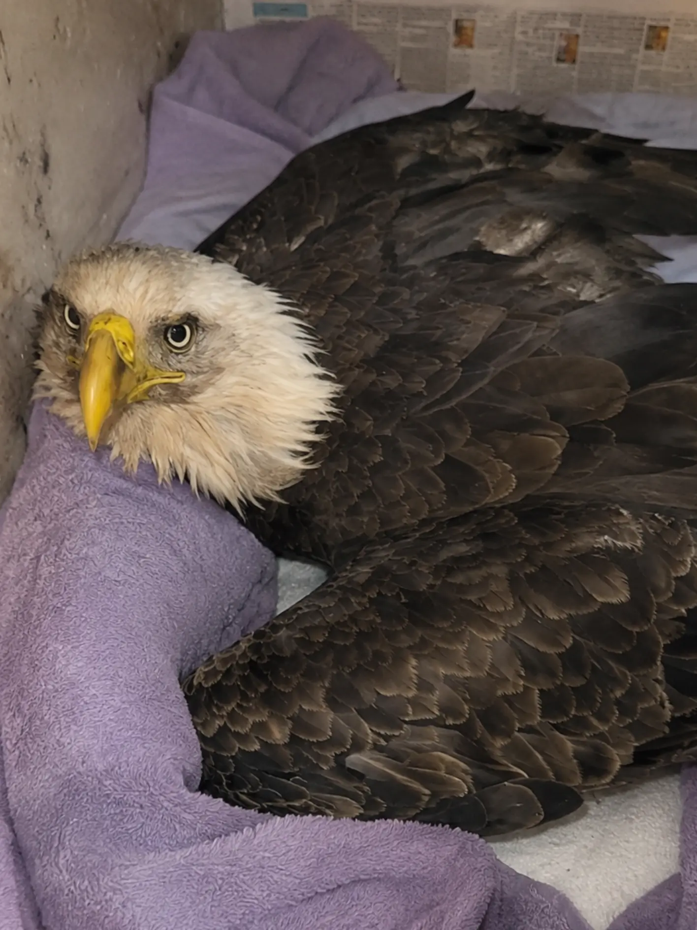
It responded to activity around it.
But every attempt to rise ended the same way.
The legs failed to support the body, and the eagle settled back onto the ground.
For an adult bald eagle — a bird capable of launching a body weighing several kilograms into the air, gripping branches in strong winds and carrying food with powerful feet — that degree of weakness was immediately alarming.
At first, trauma seemed like an obvious possibility.
A grounded eagle with weak legs could have suffered a pelvic fracture, spinal injury, damage to one or both legs, or blunt-force trauma from a collision.
But the initial examination did not reveal the kind of external injury that would normally explain complete loss of normal weight-bearing.
The bird could move its toes.
It retained some response in both feet.
There was no obviously displaced limb.
And when radiographs were obtained, veterinarians found no major fracture or dislocation severe enough to explain why the eagle could not stand.
That changed the direction of the case.
The team began looking beyond the bones.
Blood was collected.
Neurological responses were monitored.
Lead testing was included in the diagnostic workup.
The result provided the first major clue.
The eagle had a markedly elevated blood lead concentration consistent with clinically significant exposure.
The weakness that initially appeared orthopedic was instead increasingly consistent with a systemic toxicological problem affecting the bird’s neurological and muscular function.
“The unusual part was that we could not identify a fracture that explained what we were seeing,” one veterinarian involved in the case said. “The eagle was alert, but the legs simply were not supporting the body normally. Once the lead result came back, the clinical picture made much more sense.”
Treatment began immediately.
The eagle was kept on padded flooring because it could not perch safely.
Fluids and nutritional support were provided as needed.
Chelation therapy was initiated to help remove circulating lead from the body.
And the rehabilitation team began watching for something that sounded deceptively simple but would become one of the most important measures of recovery:
Could the eagle stand again?
Found Grounded in Skagit County
The adult eagle was discovered near agricultural and wetland habitat outside Mount Vernon in Skagit County.
The region is well known for wintering waterfowl, river systems, open farmland and abundant raptor activity.
Bald eagles move through the area regularly.
They may feed on fish, waterfowl, carrion and other available food sources.
Seeing an eagle in the landscape is normal.
Seeing one sitting low on the ground and failing to leave is not.
The person who first noticed the bird initially waited at a distance to determine whether it was simply resting.
It was not.
When the eagle attempted to reposition itself, the weakness became apparent.
Instead of rising fully onto both feet, the body remained low.
The legs appeared unable to sustain weight for more than a brief period.
The bird did not walk away.
It did not take flight.
A wildlife responder was contacted.
The Eagle Was Alert but Functionally Grounded
One of the more confusing aspects of the case was the contrast between the eagle’s mental alertness and its physical weakness.
The bird watched the responder.
Its eyes tracked movement.
It showed awareness of the surroundings.
But it could not use the lower body normally.
That distinction mattered.
A profoundly unconscious bird suggests a very different set of problems from an alert eagle whose legs repeatedly collapse.
The responder approached cautiously.
Weak does not mean harmless.
The talons of a bald eagle can remain dangerous even when the bird cannot stand normally.
The feet were controlled first.
The wings were supported against the body.
The eagle was placed in a padded transport carrier and taken for veterinary evaluation.
The Intake Image Shows an Alert Bird That Could Not Support Itself Normally
Images from the early phase of care show the adult eagle resting low on towels inside a medical enclosure.
The white head is upright.
The eyes are open and attentive.
The wings remain folded against the body.
But the bird is not standing.
Most of its weight is resting directly on the padded floor.
That posture became one of the defining features of the case.
There was no dramatic open wound.
No obviously shattered wing.
No visible leg deformity.
The primary abnormality was function.
A bird built to stand, perch and launch into flight could not support its own body.
The Examination Began With the Legs
Veterinary staff first examined the structures most obviously involved.
Both legs were palpated.
The hips and pelvis were assessed.
The feet were examined for swelling, wounds, abnormal position or fractures.
The toes were checked individually.
Staff evaluated whether the eagle could flex and extend them and whether normal withdrawal responses were present.
The bird still had some movement.
That was encouraging.
But grip strength was weak.
When the body was supported and the feet were placed beneath it, the eagle could not maintain a normal standing posture for long.
The weakness appeared bilateral rather than isolated to one leg.
That observation was important.
A fracture generally creates a more localized problem.
A bird with one broken leg may shift weight aggressively onto the opposite side.
This eagle seemed globally weak in both lower limbs.
That pattern raised concern for a neurological, metabolic or toxicological problem.
Wings and Spine Were Also Evaluated
The veterinary team could not assume the problem stopped at the legs.
The shoulders were checked.
Both wings were extended carefully.
Major wing bones were palpated.
The spine and pelvis were assessed for signs of trauma.
The head and neck were also examined.
No major external injury emerged that could fully explain the inability to stand.
Because some skeletal injuries remain hidden beneath feathers, radiographs were the next step.
X-Rays Ruled Out One of the Biggest Concerns
Radiographs were obtained to look for:
- fractures of either leg,
- pelvic injury,
- hip abnormality,
- spinal trauma visible on standard imaging,
- and other skeletal damage.
The images did not reveal a major fracture or dislocation.
That was good news.
But it also deepened the diagnostic question.
If the bones were intact, why was the eagle unable to walk?
The team now had to consider diseases that interfere with muscle control, nerve function or systemic strength.
Neurological Evaluation Became Central
Clinicians began paying close attention to the bird’s neurological responses.
Was the head held normally?
Were the pupils symmetrical?
Did both eyes respond appropriately?
Was there a head tilt?
Could the bird right itself?
Were the toes being placed normally?
Did each foot withdraw from controlled stimulation?
Was grip symmetrical?
The eagle remained mentally responsive.
There was no dramatic progressive head tilt or obvious seizure activity at that stage.
The most prominent abnormality remained weakness in the legs and feet.
That narrowed the problem without solving it.
Bloodwork Was Collected
The rehabilitation center collected blood for a broader medical evaluation.
Routine testing could help identify:
- anemia,
- dehydration,
- inflammation,
- metabolic abnormalities,
- or evidence of organ dysfunction.
For a grounded bald eagle with unexplained weakness, lead screening was also considered clinically important.
Bald eagles are opportunistic scavengers.
They may consume carrion or animal remains containing small fragments of lead ammunition.
Those fragments can be difficult or impossible to see in tissue.
An eagle does not need to swallow an intact bullet to be exposed.
Tiny particles may be enough to introduce lead into the gastrointestinal tract.
Once absorbed, lead can affect multiple systems.
The Lead Result Changed the Case
The blood lead concentration returned markedly elevated.
That finding provided a plausible explanation for the weakness.
Lead toxicosis can affect the nervous system.
It can interfere with normal muscle coordination.
It can contribute to gastrointestinal dysfunction.
It can alter red blood cell production.
Affected birds may become weak, unable to perch normally, unable to fly, poorly coordinated or increasingly neurologically abnormal.
In this eagle, the most striking sign was the loss of normal lower-body strength.
The bird remained aware of its surroundings.
But the neuromuscular system was not functioning normally enough to support standing and walking.
“The lead result gave us an explanation that the radiographs could not,” the veterinarian said. “The bird’s legs were not broken. The problem was that the system controlling normal strength and coordination was compromised.”
The Exact Source of Lead Could Not Be Identified
Testing can show that a bird has been exposed.
It usually cannot identify where the exposure occurred.
The eagle may have scavenged on an animal carcass containing lead fragments.
It may have consumed contaminated tissue elsewhere in the landscape.
The exposure could have occurred days before the bird was found.
No single carcass or feeding event was identified.
That uncertainty is common.
Bald eagles travel widely.
They may move between rivers, agricultural land, road corridors and open fields while feeding opportunistically.
By the time clinical signs become severe enough for a bird to be grounded, the original source may be impossible to reconstruct.
The rehabilitation team therefore avoided assigning a specific source without evidence.
Treatment Focused First on Stabilization
Before neurological recovery could be evaluated, the eagle had to be supported safely.
Because it could not stand reliably, the bird was housed on padded flooring.
High perches were removed.
The enclosure was kept quiet.
Handling was minimized.
Towels and soft bedding helped protect the breastbone, feet and feathers from prolonged pressure.
A bird designed to spend much of its time standing or perching can develop secondary problems if forced to remain recumbent for long periods.
Staff therefore monitored the eagle’s body position carefully.
Fluid Therapy Supported the Patient
If dehydration or poor intake was present, fluid support could be used to stabilize circulation and help the body maintain normal organ function.
Fluids do not remove lead directly.
They support the patient while specific treatment addresses the toxic exposure.
That difference is important.
A poisoned eagle may require several forms of care at once.
Chelation addresses lead.
Fluids support hydration.
Nutrition maintains energy.
Padded housing prevents secondary injury.
Monitoring identifies whether neurological function is returning.
The treatment plan has to address the entire animal.
Chelation Therapy Was Initiated
Because the blood lead concentration was considered clinically significant in the context of the eagle’s weakness, chelation therapy was initiated.
Chelating medications bind to lead and help the body eliminate it.
But chelation is not an instant antidote.
Removing lead from circulation does not guarantee that neurological function will return immediately.
Some damage may take time to resolve.
The nervous system has to recover.
Muscles weakened by days of poor function may need to rebuild.
The bird may continue appearing severely debilitated even while the blood lead concentration begins moving in the right direction.
That is why clinicians watch both laboratory values and behavior.
Lower Lead Levels Alone Would Not Be Enough
A repeat blood result showing improvement would be encouraging.
It would not prove the eagle was ready for release.
The rehabilitation team needed the bird to regain function.
Could it rise?
Could it stand?
Could it grip?
Could it perch?
Could it walk or reposition normally?
Eventually, could it fly and land without collapsing?
Those functional milestones would matter just as much as the laboratory number.
“A lower blood lead level is good news,” the veterinarian said. “But this bird has to do everything a wild eagle needs to do before release. It has to stand, perch, grip, fly and land normally. We cannot make that decision from one blood test.”
Nutrition Was Closely Monitored
Weak raptors may eat poorly.
They may also struggle to manipulate food when they cannot stand normally.
The eagle’s appetite was therefore monitored carefully.
Food was positioned so the bird could access it without having to balance on a high perch.
If voluntary intake was insufficient, nutritional support could be provided cautiously.
Maintaining body condition was essential.
The bird needed energy for neurological recovery, muscle maintenance and general healing.
Prolonged recumbency combined with poor food intake could rapidly create a second problem: severe deconditioning.
The Feet Were Protected During Recumbency
Because the eagle remained low for much of the early hospitalization, staff watched the feet closely.
Pressure injuries can develop in captive raptors when weight is distributed abnormally for extended periods.
Bedding was kept clean and dry.
The eagle was given enough support to remain comfortable without being forced to stand.
As strength returned, natural repositioning was encouraged rather than constant manual manipulation.
The rehabilitation team wanted movement to come from the eagle itself whenever possible.
That would provide the best evidence that neurological control was returning.
The First Improvement Was in the Grip
Recovery did not begin with flight.
It began in the toes.
After treatment had been underway, staff noticed that the eagle’s grip was becoming firmer.
The feet responded more strongly when placed against a stable surface.
Toe flexion became more purposeful.