When a Bald Eagle was first brought into River Valley Veterinary Hospital after being found near Lake Pepin in Wabasha County, Minnesota, there was one detail that initially gave rescuers reason for hope.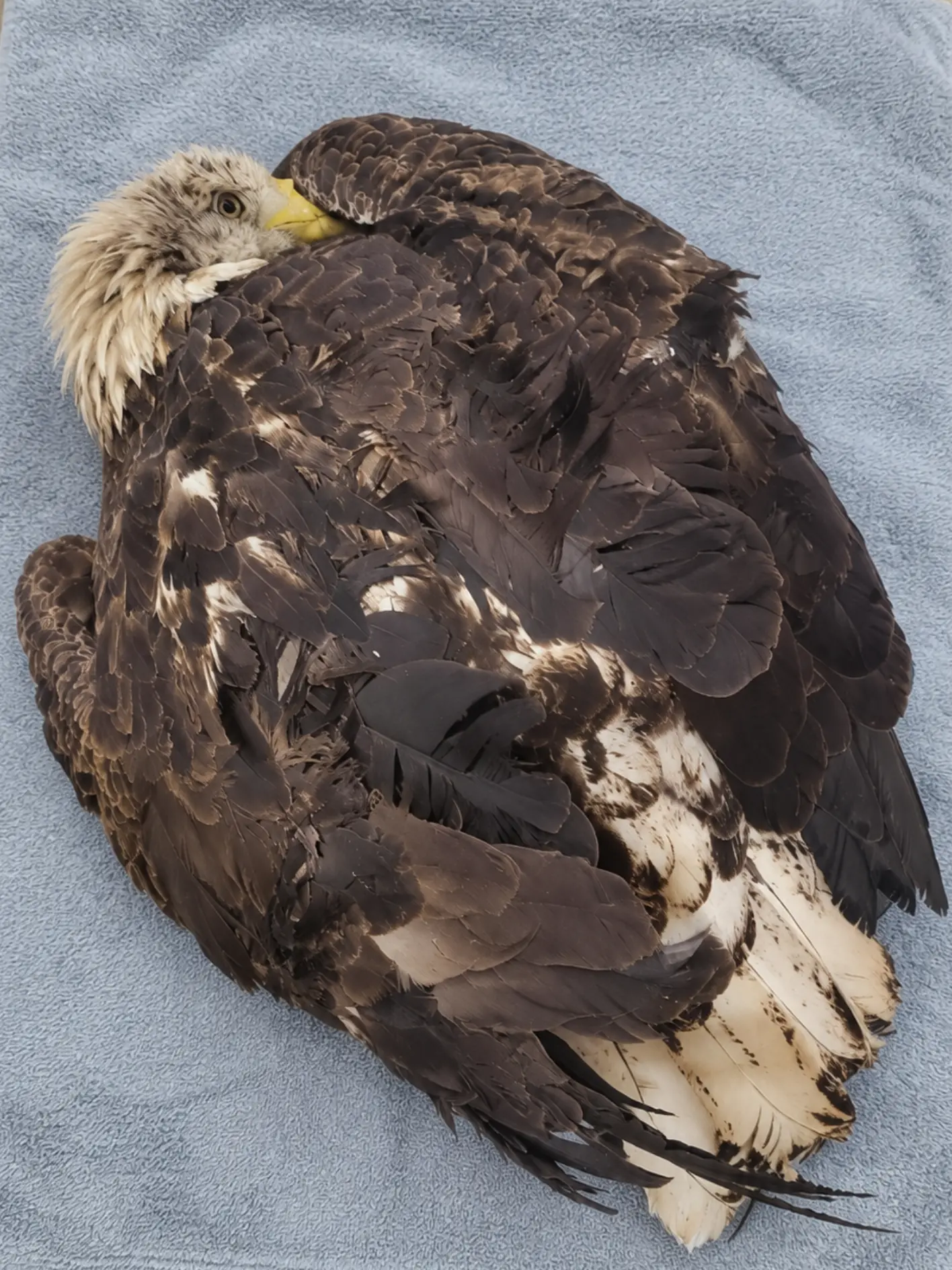
The eagle could still stand.
It was weak and unusually tolerant of people, and it had not flown when approached, but both wings appeared intact. There was no exposed bone, no major external bleeding, and no immediately obvious injury severe enough to explain why a wild adult eagle had remained on the ground.
Conservation Officer Mark Jensen, who transported the bird from the scene, initially suspected some form of trauma. A vehicle collision, an unseen wing injury, or a hard impact remained possible.
Radiographs were obtained.
They showed no broken bones.
For a moment, that seemed like good news.
But it also removed one of the simplest explanations for the eagle’s condition.
“The X-rays told us what wasn’t wrong,” one member of the veterinary team said. “They didn’t tell us why this bird couldn’t fly.”
Within hours, the eagle’s condition began to deteriorate.
The bird that had been able to stand at admission started crouching lower. Its grip weakened. It became increasingly lethargic. Eventually, it could no longer support its own weight.
That change shifted the concern away from an isolated orthopedic injury and toward a systemic or neurologic problem.
A blood sample would ultimately provide the answer.
The first attempt to measure the eagle’s lead concentration exceeded the reportable range of the analyzer being used. The sample had to be diluted and tested again.
The final result was extraordinary:
261 micrograms of lead per deciliter of blood.
The eagle did not have a broken wing.
Its body was being overwhelmed by lead.
Found Near Lake Pepin
The eagle had first been reported after a resident noticed it sitting unusually low near the edge of the Lake Pepin area.
Bald Eagles are common along portions of the Upper Mississippi River system, where open water, fish, waterfowl, and carrion provide abundant feeding opportunities. Seeing one on the ground is not necessarily an emergency.
What concerned the observer was the bird’s behavior.
It remained in place.
It did not fly when people moved within a distance that would normally make a healthy adult eagle uncomfortable.
Its head was upright, and it was visually responsive, but its reactions seemed slow.
When Jensen arrived, the eagle was still capable of supporting itself on both feet.
That mattered.
A severely injured raptor may collapse immediately. This bird had enough muscular control to remain upright, and its wings were folded normally against the body.
But it would not fly.
Jensen approached carefully.
Even sick Bald Eagles can suddenly defend themselves with their feet, and a bird that appears nearly motionless can still strike with considerable force.
Once the talons were safely controlled, the eagle was placed into a padded transport carrier and taken to River Valley Veterinary Hospital.
No attempt was made to force the bird into flight.
That decision would prove especially important once the true diagnosis became clear.
X-Rays Found Nothing Broken
At the hospital, veterinarians performed a physical examination and obtained radiographs of the wings, body, and other relevant structures.
The team was specifically looking for injuries that might explain the eagle’s inability to leave the ground.
No major wing fracture was found.
The radius and ulna were intact.
There was no obvious pelvic fracture.
No catastrophic spinal injury was identified.
The skeletal structures that should have allowed the bird to stand and fly were largely intact.
That was encouraging from an orthopedic standpoint.
But clinically, it created a more troubling question.
If the bones were not broken, why was the eagle behaving as though its body could no longer use them properly?
The answer began emerging as the bird’s condition worsened.
The Eagle Started Losing the Ability to Stand
Initially, the eagle could remain upright.
Then it began spending more time crouched.
Its feet still moved, but the grip was weaker than expected from an adult Bald Eagle.
The bird became progressively less responsive.
Its head lowered more frequently.
By the time the decision was made to transfer the eagle for specialized wildlife care, it could no longer stand reliably.
That progression was a significant diagnostic clue.
Traumatic injuries generally do not disappear on X-rays while causing rapidly worsening, symmetrical weakness without some other evidence of damage. A toxicologic, metabolic, or neurologic problem became increasingly likely.
Lead exposure immediately moved higher on the list.
Bald Eagles are particularly vulnerable because of their feeding ecology.
They are highly capable predators, but they are also scavengers.
A free-ranging eagle may consume deer remains, wounded game animals, discarded internal organs, or other carcasses encountered across its territory.
If those tissues contain fragments of lead ammunition, an eagle may ingest the metal unintentionally.
The bird does not need to be shot.
It only needs to eat contaminated tissue.
A Transport Relay Was Organized
River Valley Veterinary Hospital could provide initial assessment and stabilization, but the eagle needed a facility experienced in long-term raptor toxicology and rehabilitation.
A transfer was arranged.
Volunteer transporters Megan Foster and Caleb Reed collected the eagle and began the next stage of the journey.
They transported the bird to Upper Midwest Wildlife Rescue, where they met transport coordinator Ruth Ellis.
Ellis then continued the final leg to Northland Raptor Medical Center, where a team prepared to receive a deteriorating eagle with no obvious orthopedic explanation for its weakness.
It was the kind of wildlife transport system that often operates almost invisibly.
One person finds the bird.
A conservation officer moves it to initial care.
A veterinary hospital stabilizes it.
Volunteer drivers take over.
Another coordinator completes the relay.
A specialty center finally begins definitive treatment.
At every stage, delay can matter.
For this eagle, the decline was already underway.
By the Time It Reached Specialty Care, It Was No Longer Standing
When the eagle arrived at Northland Raptor Medical Center, the difference from its first examination was striking.
The bird was now recumbent.
Instead of standing with its feet beneath the body, it remained curled low on soft bedding.
A photograph taken during this stage shows the eagle resting on a blue towel, one eye still open but the entire body held close to the surface.
There is no visible catastrophic injury.
That is precisely what made the case so deceptive.
A viewer might see an exhausted bird.
The clinicians saw neurological weakness.
The eagle had structurally intact wings and legs.
What it was losing was the ability to control and use them normally.
The medical team moved quickly.
A blood sample was collected almost immediately.
The First Lead Test Could Not Produce a Final Number
Lead screening is an important part of the evaluation for Bald Eagles presenting with unexplained weakness, inability to fly, reduced grip, abnormal posture, or neurological impairment.
The first analysis produced an unusual result.
The concentration was above the instrument’s direct reportable range.
In practical terms, the machine could tell the team that the sample contained a very high concentration of lead, but it could not provide the final quantitative result using the initial preparation.
The sample had to be diluted and run again.
“The first result did not give us a usable final number,” a clinician said. “It told us that the lead concentration was beyond the range we could report directly. At that point we knew we were dealing with a very serious exposure.”
The diluted sample was analyzed.
This time, the laboratory obtained a result:
261 µg/dL.
That number finally explained what the radiographs had not.
The eagle was not grounded because its bones had failed.
Lead was interfering with the biological systems that allowed those intact bones, muscles, and nerves to work together.
A Blood Lead Level of 261 µg/dL Represented a Severe Toxicologic Emergency
The veterinary team interpreted the result as profoundly elevated and clinically dangerous.
Lead can affect multiple organ systems in birds.
The nervous system is particularly important in severely affected raptors.
Clinical signs can include weakness, reduced coordination, abnormal posture, poor grip, inability to stand, inability to fly, altered mental status, and, in advanced cases, more severe neurologic dysfunction.
The gastrointestinal system may also be affected.
Appetite can decline.
Birds may lose weight.
Generalized debilitation follows.
A Bald Eagle does not need to reach complete paralysis for lead poisoning to become life-threatening.
Even partial impairment can disrupt the precision required for survival.
A healthy eagle must coordinate vision, balance, wing movement, and foot placement continuously.
It must respond to hazards within seconds.
If those neurological processes begin to slow or fail, normal behaviors can become impossible.
The fact that this eagle had been able to stand earlier did not contradict the diagnosis.
It demonstrated something wildlife clinicians frequently see:
wild animals can appear functional until physiologic reserve begins to disappear.
Why Could the Eagle Stand at First?
Lead poisoning does not always progress in a simple, predictable sequence.
Two birds with significant exposure may present very differently.
One may arrive completely recumbent.
Another may still stand, watch handlers, and even attempt to defend itself before deteriorating.
Free-ranging wildlife also tends to conceal weakness.
A Bald Eagle surviving outside cannot afford to advertise vulnerability.
By the time obvious neurological deficits appear, the underlying problem may already be advanced.
That may explain why this bird initially appeared stronger than its bloodwork suggested.
It had not yet lost every motor function.
But the toxic process was already severe.
Within hours, the ability to remain upright began disappearing.
“The fact that the eagle stood when it first came in did not mean the nervous system was normal,” a veterinarian explained. “It meant we were watching the disease progress.”
Where Did So Much Lead Come From?
The precise source could not be identified.
That is common in free-ranging wildlife.
A Bald Eagle may travel across a wide territory and feed at many locations before becoming ill enough to be found.
There is usually no way to trace one individual bird back to one specific carcass.
But the exposure pathway is well established.
Bald Eagles frequently scavenge animal remains.
When animals are shot with lead ammunition, projectiles can fragment on impact.
Some particles remain near the wound channel.
Others may disperse through surrounding tissue.
Fragments can also remain in discarded internal organs or carcasses left accessible to scavengers.
The pieces do not need to look like bullets.
Some are extremely small.
An eagle feeding on meat can swallow them unknowingly.
Inside the digestive tract, metallic lead can become available for absorption.
Once absorbed, it enters the bloodstream and can affect tissues throughout the body.
The veterinary team could not say which carcass had exposed this particular eagle.
But ingestion of lead ammunition fragments in scavenged animal remains was a medically plausible and well-recognized route.
Treatment Could Not Wait
At 261 µg/dL, the case was treated as a serious toxicologic emergency.
The medical team began chelation therapy, using medication designed to bind lead and help facilitate its elimination from the body.
The eagle also needed intensive supportive care.
It was dehydrated.
It could not stand.
Its neurological function was impaired.
Food intake had to be monitored carefully.
The bird was placed on soft, padded flooring rather than a perch.
That was essential.
A neurologically weak eagle can fall and create an entirely new injury if placed above the ground too early.
Rolled towels and soft bedding helped keep the body in a safer position.
Staff monitored pressure points, breathing, responsiveness, foot movement, appetite, hydration, and body condition.
The first objective was not flight.
It was survival.
Why the Bird in the Photograph Is Lying Down
A Bald Eagle normally spends much of its time standing or perched.
Seeing one lying low on towels is therefore clinically significant.
In this case, the bird did not have a fractured leg preventing it from standing.
Instead, systemic weakness and neurological dysfunction had left it unable to support normal posture safely.
The padded environment served several purposes.
It reduced the risk of injury during uncontrolled movements.
It protected the feet and keel.
It allowed the eagle to rest without falling from a perch.
And it gave staff an easier way to monitor changes in mobility.
Something as small as a toe movement could become meaningful.
The First Night Remained Uncertain
Despite treatment, no one could guarantee that the eagle would survive.
The lead concentration was extremely high.
The bird was profoundly weak.
Its inability to stand indicated that the toxic exposure was no longer merely a laboratory finding.
There were clear clinical consequences.
The eagle rested for long periods.
Its eyes opened intermittently.
Grip remained poor.
Appetite was limited.
The veterinary team monitored the bird closely while chelation and supportive care continued.
The prognosis remained guarded.
The people who had transported the eagle waited for updates.
Megan, Caleb, Ruth, and Jensen had all handled only one section of the case.
None of them knew whether they had moved a future release candidate or an eagle in the final hours of its life.
Eating Became the First Encouraging Sign
Then the eagle showed interest in food.
At first, the change was modest.
The bird became more alert when food was presented.
Later, it began eating voluntarily.
For the rehabilitation team, that was significant.
A bird feeding independently must coordinate vision, head movement, beak function, swallowing, and enough mental awareness to recognize and respond to food.
“Eating was the first sign that the eagle was beginning to reclaim something the lead had taken away,” one rehabilitator said.
The bird was still unable to stand normally.
No one considered it out of danger.
But the direction had changed.
For the first time since the dramatic decline began, the team was seeing function return rather than disappear.
The Lead Level Began to Fall
Blood samples were repeated during treatment.
The lead concentration declined.
That was encouraging, but the veterinary team did not rely on one lower number.
Lead toxicosis requires clinical monitoring as well as laboratory monitoring.
A bird can have an improving blood concentration and still retain neurological deficits.
Conversely, a bird can look brighter before the lead burden is sufficiently controlled.
The team continued asking practical questions.
Could the eagle grip?
Could it hold its head normally?
Could it reposition itself?
Would it attempt to stand?
Was appetite consistent?
Did mental responsiveness remain appropriate?
Those answers would ultimately matter as much as the laboratory number.
The First Neurological Milestone Came From the Feet
For a patient whose eventual goal is flight, the earliest meaningful recovery can look remarkably small.
One day, the eagle closed its toes more firmly around a rolled towel.
The grip was still weak compared with that of a healthy adult.
But it was purposeful.
That mattered.
Normal foot function requires communication between the brain, spinal pathways, peripheral nerves, and muscles.
A stronger grip suggested that some neurological control was returning.
Over subsequent examinations, the toes became more responsive.
The eagle began pushing against the bedding.
Its body rose slightly higher.
The first major sign of recovery was not a wingbeat.
It was a foot closing around fabric.
Standing Had to Be Relearned Gradually
The eagle’s recovery progressed in stages.
First, it remained lying low.
Then it began maintaining a more upright resting posture.
Later, it pushed itself onto its feet for brief periods.
The first attempts did not last long.
Weakness forced the bird down again.
Staff did not push it to remain standing.
A recovering neurological patient can fatigue quickly, and repeated falls would create unnecessary trauma.
Instead, progress was allowed to come gradually.
Seconds of weight-bearing became longer periods.
The feet began positioning more normally.
Balance improved.
Eventually, the eagle could stand without immediate collapse.
That was a major milestone.
It was also only the beginning.
A Lower Blood Lead Level Did Not Automatically Mean Recovery
One of the most important principles in this case was that detoxification and functional recovery were not the same thing.
Chelation could lower circulating lead.
It could not guarantee that every neurological effect would immediately disappear.
Some lead-exposed birds can retain weakness, coordination deficits, or other abnormalities even after blood values improve.
For a captive bird, mild impairment might be manageable.
For a wild Bald Eagle, mild impairment can be fatal.
That meant this eagle could not be considered releasable simply because laboratory values improved.
The team needed evidence that the nervous system was working normally again.
A Low Perch Became the Next Test
Once standing was consistent and grip strength improved, the eagle was given access to a low perch.
The height was intentionally conservative.
Perching tests several systems simultaneously.
The toes must grip.
The legs must support body weight.
Balance must remain stable.
Vision must help guide body position.
The nervous system must make continuous small adjustments.
At first, the eagle shifted frequently.
Then it became steadier.
The feet remained closed securely around the perch.
Head position stayed normal.
The bird began behaving increasingly like a raptor rather than a critical-care patient.
Staff also noticed another encouraging development.
The eagle was becoming more defensive.
Handling was getting harder.
That was good.
Wild Behavior Was Returning
A healthy adult Bald Eagle should not be comfortable with human handling.
As strength returned, the bird began objecting more forcefully during examinations.
Its eyes followed staff immediately.
The feet became increasingly dangerous again.
The eagle tried to create distance whenever possible.
Wildlife rehabilitators welcomed those behaviors.
The purpose of treatment was not to tame the bird.
It was to restore independence.
The more the eagle behaved like something that wanted nothing to do with people, the closer it moved toward that objective.
Flight Would Be the Most Difficult Neurological Test
Eventually, standing and perching were no longer enough.
The eagle had originally been rescued because it could not fly normally.
Before release could even be discussed, the bird would need to prove that the nervous system could manage one of the most demanding behaviors in avian biology.
Flight requires far more than functional wings.
The brain must process visual information rapidly.
Both wings must move symmetrically.
The tail must help control direction.
The bird must respond to changes in speed and position.
The feet must extend at precisely the correct moment before landing.
Balance must remain stable throughout the sequence.
A subtle neurological deficit that is almost invisible on a hospital floor can become obvious in the air.
That is why the eagle was not rushed into a flight enclosure.
The First Flight Was Only the Beginning
When clinicians were satisfied with the eagle’s standing, perching, body condition, and neurological status, it was moved into a larger rehabilitation enclosure.
Staff stepped back.
The eagle was allowed to choose when to move.
Eventually, it launched.
Both wings extended.
The bird crossed the enclosure and reached another perch.
The flight was controlled.
That was an enormous improvement over the animal that had been unable to stand.
But one successful flight was not enough.
The team needed repetition.
Could the eagle do it again?
Could it turn?
Could it gain altitude?
Could it land accurately?
Would fatigue reveal neurological weakness?
Those questions required additional time.
Repeated Flights Provided the Evidence
Over subsequent rehabilitation sessions, the eagle continued flying.
Strength improved.
Turns became more precise.
The bird reached different perch heights.
Landings remained controlled.
No obvious neurological deficits appeared after exertion.
Grip remained strong.
Appetite was normal.
Body condition improved.
Repeat lead testing continued to support the clinical recovery.
Gradually, the case moved from a toxicologic emergency toward a release assessment.
The people involved in the original transport began receiving very different updates.
The eagle was standing.
Then perching.
Then flying.
For a bird they once feared might die somewhere between facilities, each message carried significance.
The Case Shows Why a “Normal” X-Ray Does Not End the Investigation
One of the clearest lessons from this eagle’s hospitalization came from the first set of radiographs.
There were no broken bones.
That did not mean there was no emergency.
A grounded eagle with intact wings and legs may still be profoundly ill.
Toxic exposure can remove function without destroying anatomy.
The bones can be normal.
The joints can be intact.
The feathers can look undamaged.
And yet the bird may be incapable of flying because its nervous system can no longer coordinate the body effectively.
That is why a full diagnostic assessment matters.
The X-ray answered one question.
The blood sample answered the more important one.
The Number 261 Explained the Collapse
The final diluted result — 261 µg/dL — became the defining measurement in the case.
It transformed unexplained weakness into a severe lead toxicosis diagnosis.
The number also explained the strange sequence rescuers had witnessed.
An eagle still able to stand.
No fractures.
Then progressive weakness.
Then inability to remain upright.
The problem had been hidden inside the bloodstream.
The bird did not need a cast.
It needed detoxification, intensive nursing care, time, nutrition, and neurological recovery.
The Source Remained Unknown, but the Prevention Message Was Clear
The medical team could not identify the specific source of this eagle’s exposure.
Responsible reporting requires acknowledging that uncertainty.
But there is a broader, well-recognized risk that can be reduced.
Lead ammunition fragments in scavenged animal remains can expose Bald Eagles and other wildlife.
Using effective non-lead ammunition where appropriate reduces that source of toxic metal in carcass tissue.
Responsible handling of potentially contaminated animal remains can also reduce opportunities for scavengers to ingest fragments.
The issue is not lawful hunting itself.
Hunters have long contributed to wildlife conservation, habitat management, and population monitoring.
The concern is the toxic material that can remain after a shot.
Lead-free alternatives can allow hunting and scavenger conservation to coexist.
Transport Volunteers Were Part of the Medical Chain
There is another lesson in this case that has nothing to do with toxicology.
The advanced hospital could not help an eagle that never reached it.
Mark Jensen brought the bird from the field.
Megan Foster and Caleb Reed carried it to the next facility.
Ruth Ellis completed the final transfer.
The veterinary team then took over.
Four people, multiple facilities, and one uninterrupted chain of care turned a grounded bird into a patient with a chance.
Wildlife rehabilitation often depends on precisely that kind of cooperation.
The medicine may happen inside an examination room.
The opportunity for medicine often begins on the road.
From Standing to Collapse — and Then Back Again
The eagle’s clinical course came full circle.
At the beginning, it could stand.
Then lead poisoning took that ability away.
The bird became recumbent.
Its grip weakened.
Its appetite declined.
A blood sample exceeded the analyzer’s initial reportable range.
The diluted result came back at 261 µg/dL.
Chelation therapy began.
The eagle ate.
The toes started gripping again.
It pushed itself upright.
It stood.
It perched.
Eventually, it flew.
That progression demonstrates why wildlife rehabilitation cannot be judged from one photograph or one laboratory result.
A bird may look stronger than it is.
A bird that looks nearly lost may still recover.
The important question is what objective function returns over time.
What the Volunteers Remember
The transporters never saw the full medical recovery while it was happening.
Their part of the story was far less dramatic.
A carrier.
A vehicle.
A handoff.
Another drive.
But they remembered how weak the eagle had become.
They remembered that it had initially stood and then deteriorated.
They remembered not knowing whether they were moving an animal toward rehabilitation or simply giving specialists a chance to understand why it was dying.
When the update finally came that the eagle was eating and becoming more alert, that alone felt significant.
Later came the message that it had begun standing.
Then perching.
Then flight rehabilitation.
The story they had expected to end during transport was continuing.
The Final Standard Was Independence
Before any release could occur, the eagle had to satisfy standards far higher than merely surviving lead poisoning.
It needed normal blood lead status appropriate for discharge.
Normal neurological responses.
Powerful bilateral grip.
Appropriate vision and awareness.
Strong body condition.
Independent feeding.
Consistent flight.
Controlled turns.
Accurate landings.
Normal endurance.
And, importantly, appropriate avoidance of people.
Only when all of those functions were demonstrated repeatedly could clinicians consider the bird capable of returning to an environment where there would be no second chance after a poorly controlled landing or delayed reaction near traffic.
That is the difference between treatment and wildlife rehabilitation.
Treatment keeps the animal alive.
Rehabilitation determines whether it can live wild again.
The Hidden Emergency Was in the Blood
When this Bald Eagle first reached River Valley Veterinary Hospital, the obvious signs were confusing.
It could stand.
Its wings were intact.
X-rays showed no fractures.
There was no massive wound.
Then, over the following hours, the bird lost the ability to support itself.
A blood sample finally revealed the reason.
The concentration of lead was so high that the original test exceeded the analyzer’s direct range and the sample had to be diluted.
The final value was 261 micrograms per deciliter.
That result explained what the X-rays could not.
Nothing essential had to be broken for the eagle to become grounded.
Lead had disrupted the systems that allowed an otherwise intact body to function.
The bird’s recovery therefore began not with orthopedic repair, but with chelation, fluids, nutrition, padded care, repeated blood testing, and patience.
The first signs were small.
An eye staying open.
An independent meal.
A stronger grip.
A few seconds standing.
Then a perch.
Eventually, wings.
For the people who had moved this eagle from one facility to another, that progression meant something simple:
the bird they had feared might die somewhere along the transport route was still there.
And now, instead of asking whether it would survive the night, the rehabilitation team could begin asking a very different question:
whether a Bald Eagle that once carried 261 µg/dL of lead in its blood could become strong, coordinated, and wild enough to return to the sky.