When the adult Bald Eagle arrived at the Atlantic Veterinary College in Charlottetown, the most troubling problem was not the blood visible around its head or the damaged feathers along its back. The bird could still move its wings, raise its head, and respond to people standing nearby, but it could not stand.
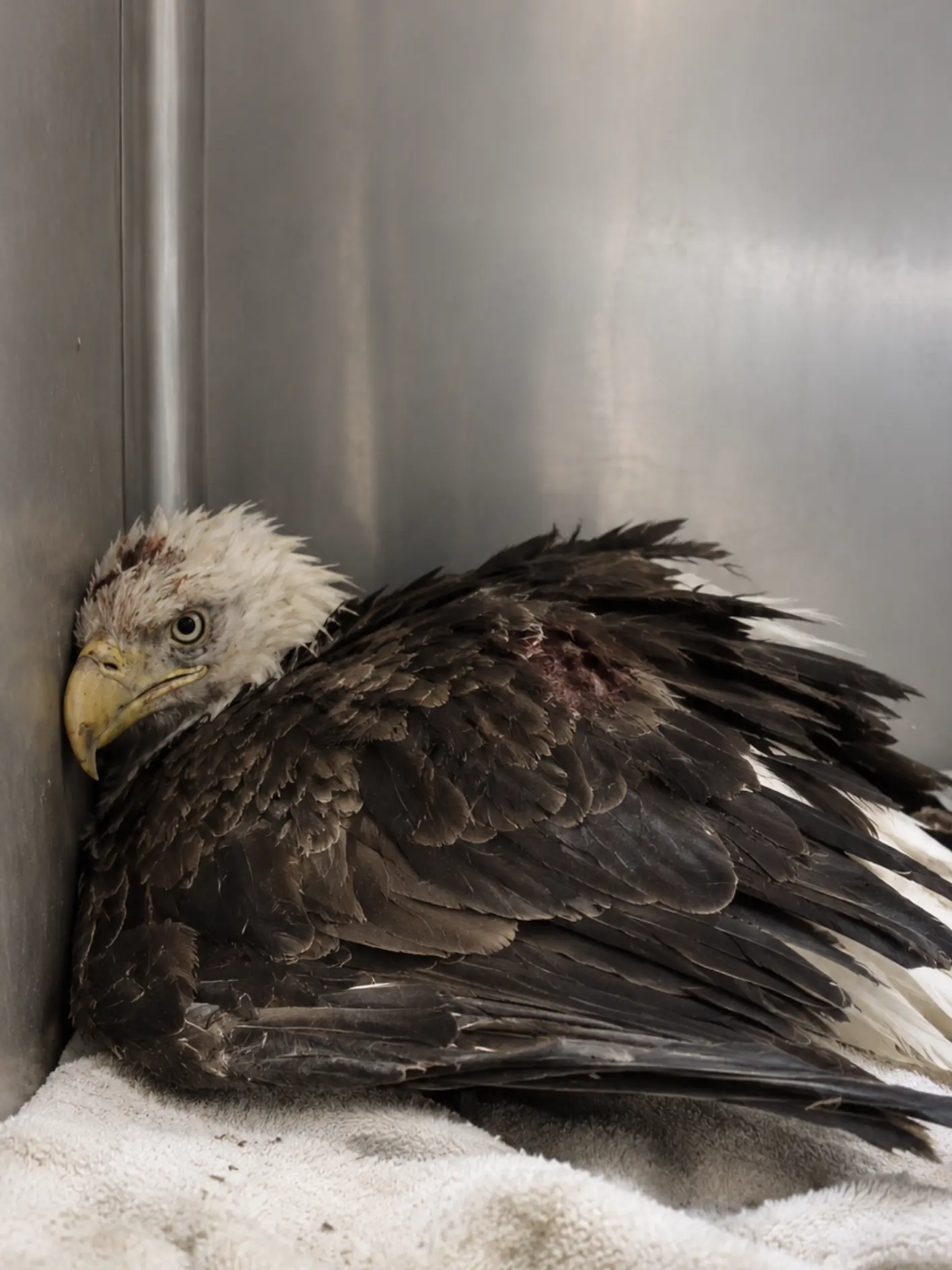
Both legs were present beneath the body, yet they were unable to support the eagle’s weight. Its feet showed only limited useful movement, and the powerful grip expected from a healthy adult Bald Eagle was markedly reduced. Whenever the bird attempted to reposition itself, it relied heavily on its wings while the lower body remained close to the padded floor.
For the veterinary team, those findings immediately raised concern about a serious neurologic injury.
The possibility of trauma to the spine or spinal cord was difficult to ignore.
Diagnostic imaging later confirmed that concern. There was a significant injury affecting a region of the vertebral column considered particularly vulnerable in large birds. The alignment of the spine had been disrupted, and the abnormality was serious enough that the medical team believed the spinal cord was being affected.
What followed was an extraordinarily difficult decision.
Conventional supportive care might allow swelling to decrease, but if the unstable vertebral injury continued compressing or damaging the spinal cord, waiting could also allow permanent neurologic deficits to become established. Surgery carried substantial risk. There was little precedent for performing this type of spinal stabilization in a free-ranging Bald Eagle, and even technically successful surgery could not guarantee that the bird would ever regain normal use of its legs.
Still, there was one reason the team believed an attempt might be justified.
The eagle could still feel its feet.
“The bird could not stand, but we were still getting neurologic responses from both legs,” said Lara Cusack, a senior clinical specialist with the wildlife service. “That did not tell us that it would recover, but it told us that the situation was not necessarily hopeless.”
Cusack also explained that there is a particular area of the spinal column that can be vulnerable to fracture in large birds such as eagles. In this case, imaging identified an injury in the region the clinicians had suspected during the neurologic examination.
The team was now faced with a question that went far beyond ordinary wound care.
Could surgeons stabilize the spine of a Bald Eagle well enough to give its spinal cord a chance to recover?
The procedure they planned may represent one of the most unusual attempts in wildlife medicine.
The Eagle’s Inability to Stand Changed the Entire Case
When wildlife patients arrive at a hospital, the first visible injury is not always the most important one.
This eagle had several external signs of trauma. There was blood around the head, disrupted feathers, and an area of damaged tissue along the upper body. Any of those findings could have justified examination on their own.
However, none of them explained why a powerful adult raptor could not rise onto its feet.
A healthy Bald Eagle has extremely strong legs and talons. Its feet are central to nearly every aspect of survival.
The bird uses them to land.
It uses them to grip branches during wind and rain.
It uses them to capture and secure prey.
It uses them to hold food while the beak tears flesh.
It depends on the legs for balance, defense, and takeoff.
An eagle that can move its wings but cannot reliably use its feet may still be incapable of surviving in the wild.
For that reason, the medical team did not interpret the condition simply as temporary weakness.
The neurologic system had to be evaluated carefully.
The First Examination Was Deliberately Conservative
The eagle was placed on thick padded towels rather than on a perch.
No one attempted to force it to stand.
Repeatedly making a neurologically compromised bird prove that it cannot walk provides little useful information and risks worsening an unstable injury.
Instead, the clinicians first evaluated respiration, circulation, body temperature, hydration, vision, and general responsiveness.
The eagle remained conscious.
Its eyes tracked movement.
The head could be held upright.
Both wings moved, although the bird appeared uncomfortable.
There was no immediate evidence that respiratory failure or massive external hemorrhage was the principal problem.
The legs remained the greatest concern.
The feet were examined individually.
The team looked for voluntary movement, muscle tone, withdrawal responses, symmetry, and evidence of sensation.
Motor function was profoundly abnormal.
The eagle could not use its legs in a coordinated way.
Yet when appropriate pressure was applied to the toes, the bird responded.
That response was repeated.
Both feet retained sensation.
It was a small finding compared with the severity of the overall case, but it became one of the most important pieces of information the clinicians had.
Preserved Sensation Did Not Mean the Bird Was Safe
Neurologic medicine is rarely simple.
The presence of sensation in a limb does not mean the animal will necessarily regain normal movement.
A spinal cord can remain partially functional while still being severely injured.
Signals may continue traveling through some neurologic pathways while others are impaired.
Inflammation, bruising, compression, and direct tissue damage can all produce different combinations of weakness and sensory loss.
The medical team therefore treated preserved sensation as an encouraging sign, not as a promise.
The eagle was still functionally unable to stand.
If that did not change, release would not be possible.
The clinicians needed to understand what was physically happening around the spinal cord.
That required imaging.
Diagnostic Imaging Confirmed the Location of the Injury
Radiographs were obtained once the bird was sufficiently stable.
The images showed abnormal alignment in a portion of the vertebral column.
There appeared to be traumatic change involving the bones that protect the spinal cord, and the position of the affected vertebrae raised concern that the neurologic structures passing through that region were being compressed or disturbed.
Radiographs are valuable for evaluating bone, but they cannot always show the spinal cord itself in detail.
For that reason, the imaging findings were interpreted together with the neurologic examination.
The two pieces of evidence pointed in the same direction.
There was a significant spinal injury.
Cusack described the affected area as one that can be particularly vulnerable in large birds.
“There is a specific region of the spine that is especially susceptible to fracture in birds of this size,” she said. “When that area is damaged, the consequences can be severe because of how closely the spinal cord is associated with the surrounding vertebral structures.”
The challenge was not simply that a bone was abnormal.
The challenge was what that abnormal bone position might be doing to the nervous system.
Birds Present Unique Challenges in Spinal Surgery
Spinal surgery is difficult in any species.
In a Bald Eagle, the anatomy makes the problem even more specialized.
Birds are built for flight.
Their skeletons must combine strength with reduced weight, and many bones differ significantly from those of mammals in shape, density, and relationship to surrounding structures.
The vertebral column also functions differently.
Certain regions are highly stabilized, while others permit movement needed for posture and flight.
That means a traumatic injury can produce a very specific pattern of instability.
The surgical team had to consider the size of the vertebrae, the limited amount of space around the spinal cord, the bird’s respiratory anatomy, positioning during anesthesia, and how any stabilization method might affect the eagle’s ability to move normally afterward.
This was not simply a smaller version of canine or human spinal surgery.
The entire operation had to be adapted to avian anatomy.
The Team Had Very Little Published Experience to Rely On
One reason the case drew such attention was the lack of established precedent.
Wildlife veterinarians routinely treat fractures of wings and legs.
They repair soft-tissue wounds.
They manage lead poisoning, infectious disease, dehydration, emaciation, and a wide range of traumatic injuries.
Spinal stabilization in a free-ranging Bald Eagle is far less common.
The team could draw on general orthopedic and neurologic principles, but there was very little published experience showing exactly how a wild eagle with this type of injury would respond.
That uncertainty affected every stage of decision-making.
Would the vertebral structures tolerate surgical stabilization?
Would the bird survive anesthesia?
Could the spine be aligned without causing additional cord injury?
Would the neurologic deficit improve afterward?
Even if the eagle eventually stood, would it regain enough grip strength to perch and land?
No one could answer those questions before surgery.
Surgery Could Stabilize the Spine, but It Could Not “Repair” the Spinal Cord
This distinction was essential.
The surgeons were not attempting to reconnect or reconstruct the spinal cord itself.
Nervous tissue does not behave like broken bone.
Instead, the goal was to repair and stabilize the damaged vertebral structures around the spinal cord.
If abnormal spinal alignment was compressing neural tissue, correcting that alignment might reduce further injury.
If the cord had been bruised or partially compressed rather than completely destroyed, stabilization could provide time for inflammation to subside and for surviving neurologic pathways to recover.
However, if major spinal cord damage had already become irreversible, no orthopedic repair could restore the missing function.
The operation could create the conditions for recovery.
It could not guarantee that recovery would occur.
The Alternative Was Equally Serious
The medical team also had to consider what would happen without surgery.
If the spine remained unstable, movement could potentially produce additional displacement.
Ongoing compression could continue affecting the spinal cord.
The eagle was already unable to stand.
If no meaningful neurologic improvement occurred, the bird would not be releasable.
Long-term quality of life would then become a major concern.
A permanently nonambulatory Bald Eagle is at risk of pressure injuries, muscle wasting, joint stiffness, chronic pain, feather damage, and significant limitations on normal behavior.
Keeping an animal alive is not the same thing as restoring an acceptable life.
Wildlife medicine has to consider both.
The team therefore weighed the uncertainty of surgery against the poor outlook if the instability remained untreated.
Ultimately, they decided the eagle had enough preserved neurologic function to justify attempting stabilization.
Preparing the Eagle for Surgery Required Careful Planning
The bird was stabilized medically before anesthesia.
Hydration was addressed.
Pain control was established.
The eagle was monitored for other injuries that could complicate surgery.
The external wounds were evaluated separately.
Bloodwork helped determine whether the bird was physiologically stable enough to tolerate a prolonged anesthetic procedure.
Only after those parameters were considered acceptable did the team move forward.
Positioning was particularly important.
The spine had to remain supported.