An adult bald eagle believed to have been struck by a vehicle while flying low across U.S. Route 101 in Mason County, Washington, is receiving intensive veterinary care after suffering a severe fracture of the humerus — one of the most important bones in the wing.
The eagle was found unable to fly near the roadway outside Shelton and was transported to a licensed wildlife rehabilitation and raptor care center in western Washington after a wildlife responder determined that the bird could not safely leave the area on its own.
What initially appeared to be a serious but potentially manageable wing fracture soon proved considerably more complicated.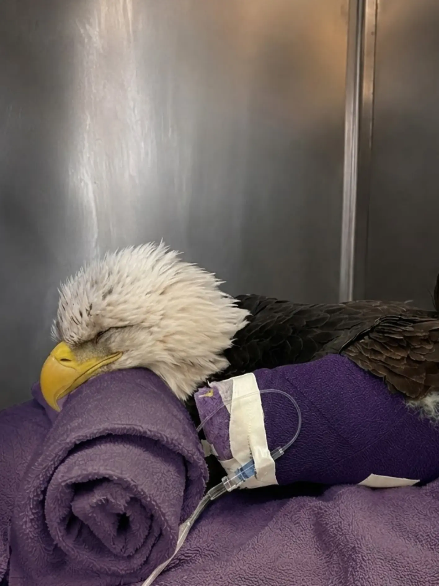
Radiographs showed that the humerus had fractured badly enough for displaced bone ends to damage the surrounding soft tissues. Veterinarians said portions of the muscle and connective tissue had been penetrated and disrupted during the traumatic event.
The medical team stabilized the wing.
They treated the open wounds.
They administered anti-inflammatory medication and antibiotics.
They provided fluid therapy.
They screened the bird for lead exposure.
They repeated radiographs to monitor healing.
They performed carefully controlled range-of-motion work while the eagle was under anesthesia.
And because the bird was not consistently eating enough on its own, staff provided assisted feeding every day to maintain body condition and support healing.
Despite those efforts, the prognosis for normal flight has continued to worsen.
The problem is no longer simply whether the broken humerus can form new bone.
Veterinarians are increasingly concerned that the combination of poor fracture geometry and irreversible soft-tissue destruction will prevent the wing from ever regaining the strength, alignment, and range of motion required for sustained flight.
“The fracture itself is only part of the injury,” one veterinarian involved in the eagle’s care said. “We can stabilize bone, treat wounds and work to preserve motion. What we cannot undo is all of the tissue damage that occurred when the fractured bone displaced through the surrounding structures.”
At this stage, the team considers the likelihood of the eagle regaining flight adequate for release to be very low.
Found Grounded Near U.S. Route 101
The eagle was discovered near a stretch of U.S. Route 101 in Mason County, an area surrounded by forest, wetlands, shoreline habitat, and waterways connected to southern Puget Sound.
Bald eagles regularly travel through the region.
They may cross roads while moving between feeding areas, waterways, roosts, and open fields.
They may also approach road corridors to investigate carrion or other food sources.
According to the circumstances reported at rescue, the bird was believed to have been struck by a vehicle while flying low near the highway.
No one was able to provide veterinarians with a complete reconstruction of the collision.
The exact speed of the vehicle was unknown.
The precise orientation of the eagle at impact was unknown.
But the combination of location, acute trauma, and the condition of the wing was consistent with a high-energy collision.
When the wildlife responder approached, the eagle remained conscious.
It could react.
But it could not achieve normal flight.
One wing was being held abnormally, and the bird remained on the ground despite the presence of people nearby.
That alone was enough to justify immediate capture.
A healthy adult bald eagle should generally be capable of creating distance from approaching humans if adequate space is available.
A bird that remains grounded after trauma may have a fracture, internal injury, neurological problem, systemic weakness, or some combination of those conditions.
The eagle was secured carefully, placed into a padded transport carrier, and taken for emergency evaluation.
The First Examination Confirmed Severe Wing Trauma
Once the bird arrived at the rehabilitation center, veterinary staff performed a complete physical examination.
The injured wing immediately drew attention.
There was swelling.
Movement was restricted.
The eagle reacted when the affected area was manipulated.
The normal anatomical relationship between portions of the wing appeared altered.
The team examined the shoulder, elbow, radius, ulna, humerus, and surrounding soft tissues.
They also evaluated the head, eyes, feet, chest, opposite wing, and general neurological status to make sure the collision had not produced additional injuries that were less obvious.
For a bird struck by a vehicle, one visible injury is rarely assumed to be the only injury.
High-energy trauma can affect multiple systems.
The eagle therefore required imaging.
Radiographs Revealed a Complex Humerus Fracture
X-rays confirmed a severe fracture of the humerus.
The humerus is the large proximal bone of the wing, extending from the shoulder toward the elbow.
Its position makes it central to normal flight mechanics.
Major flight muscles act through structures connected to the shoulder and humeral region.
A fracture there can interfere with:
- wing extension,
- wingbeat power,
- alignment,
- joint function,
- and the transfer of muscular force through the limb.
In this case, the fracture was not a simple clean break.
The bone had displaced.
The fragments were not sitting in a configuration veterinarians considered favorable for a straightforward return to full mechanical function.
More importantly, the broken ends had damaged nearby soft tissue.
That discovery changed the prognosis immediately.
A Broken Wing Bone Is Only One Part of Flight
A common misunderstanding is that a bird should be able to fly again once a broken bone has “healed.”
For large raptors, the standard is much higher.
Bone union is only one component.
A releasable bald eagle needs the entire wing to function as a highly coordinated system.
The shoulder must move through an appropriate range.
The elbow has to flex and extend.
Muscle groups must generate enough force.
Tendons and ligaments must transmit that force.
Nerves need to function normally.
Blood supply must remain adequate.
Feathers must be positioned correctly.
And the wing has to move symmetrically with the opposite side.
A bone can become radiographically stable while the wing remains mechanically inadequate for flight.
That is especially true after a complex proximal-wing fracture.
The Bone Fragments Had Damaged Surrounding Tissue
The most serious part of the eagle’s injury was what happened beyond the fracture line.
Displaced bone ends had penetrated and disrupted surrounding soft tissue.
That can cause:
- tearing of muscle fibers,
- injury to connective tissue,
- disruption of blood vessels,
- inflammation,
- contamination,
- scar formation,
- and loss of normal tissue glide.
Some of those structures can heal.
Others cannot return to their original state.
That distinction is central to the prognosis.
“The question is not simply whether the humerus develops a callus,” the veterinarian said. “The wing has to heal in a way that preserves enough muscle function and enough range of motion to produce normal flight. In this case, the tissue damage makes that much more difficult.”
The Risk of Infection Was Immediate
A complex fracture associated with soft-tissue disruption carries another concern: infection.
Once damaged bone and deeper tissues are exposed or contaminated, bacteria can enter areas that normally remain protected.
That creates the possibility of:
- deep soft-tissue infection,
- abscess formation,
- cellulitis,
- delayed wound healing,
- and potentially infection involving bone.
The veterinary team therefore treated the wounds aggressively from the beginning.
Feathers around affected areas were separated or clipped as needed to allow access.
The damaged tissue was inspected.
Wounds were flushed and cleaned.
Any visibly compromised tissue was evaluated for viability.
The goal was to reduce contamination without causing additional injury to tissue already under stress.
The Wing Was Stabilized
The fractured wing also had to be immobilized.
Every uncontrolled movement risked additional displacement.
That could cause more pain.
More tissue tearing.
More bleeding.
And further loss of alignment.
The wing was stabilized in the best achievable position based on the fracture pattern and the bird’s condition.
Stabilization does not guarantee perfect alignment.
With a complex fracture, especially one involving substantial soft-tissue trauma, the objective may initially be to prevent additional damage while allowing inflammation to settle and the patient to become physiologically stronger.
The rehabilitation team continued reassessing the wing as healing progressed.
Anti-Inflammatory Medication and Pain Control Became Part of Daily Care
Pain can interfere with nearly every aspect of recovery.
A painful eagle may refuse food.
It may resist normal posture.
It may hold the wing abnormally.
It may remain stressed even in a quiet enclosure.
Veterinary staff therefore used anti-inflammatory and pain-management therapy when clinically appropriate.
The goal was not merely to make the bird appear comfortable.
Controlling inflammation can also help reduce secondary tissue damage and allow more meaningful evaluation of wing function once the acute phase begins to improve.
However, medication cannot reconstruct destroyed muscle.
It supports the recovery process.
It does not erase the original trauma.
Antibiotics Were Used Because the Wounds Were High-Risk
Antibiotic therapy was also part of the treatment plan.
The location and severity of the wounds made bacterial infection a legitimate concern.
Again, antibiotics address only one component of the injury.
They can help control susceptible bacterial organisms.
They cannot restore damaged anatomy.
That is why even a bird that avoids infection can still have a poor flight prognosis.
The eagle’s case became a balance between preventing complications that could still be prevented and recognizing damage that medicine could not reverse.
The Image Shows the Intensity of Supportive Care
During hospitalization, the eagle has spent periods resting in a carefully supported position.
Images from treatment show the bird lying on padded towels with the body partially wrapped and supported.
Medical tubing is visible near the wrapped portion of the body, and the head rests on a rolled towel while the bird remains quiet.
The image reflects the level of supportive care required during the most difficult phase of recovery.
It should not be interpreted as a complete medical record of the procedures taking place at that exact moment.
What it does show clearly is that this was not a bird simply waiting in a cage for a fracture to heal.
The eagle required ongoing veterinary intervention.
Fluid Therapy Supported the Bird Through the Acute Phase
Trauma patients may arrive dehydrated or physiologically stressed.
Pain and reduced feeding can worsen that problem.
Fluid therapy was used to support hydration and circulation while staff treated the orthopedic and soft-tissue injuries.
Fluids do not make bone heal faster on their own.
They support the patient so normal biological healing processes can continue.
Maintaining hydration is especially important when an animal is receiving medication, coping with inflammation, and eating less than normal.
The team had to treat the entire eagle, not only the wing.
Assisted Feeding Became Necessary
One of the most labor-intensive aspects of the bird’s hospitalization has been nutrition.
The eagle did not consistently consume enough food independently during portions of its recovery.
That is not unusual in severely injured wildlife patients.
Pain.
Stress.
Immobilization.
Medication.
And general weakness can all suppress normal feeding behavior.
Staff therefore provided assisted feeding on a daily basis when needed.
The objective was to provide sufficient calories and protein to prevent further loss of body condition.
Fracture healing is metabolically expensive.
Soft-tissue repair requires energy.
Immune function requires energy.
A bird that becomes increasingly emaciated has fewer resources available for all of those processes.
Maintaining nutrition therefore became part of the orthopedic treatment even though it did not directly involve the wing.
Body Weight Was Monitored Closely
The eagle’s weight and muscle condition were tracked over time.
Veterinarians were particularly interested in the muscles along the keel.
A bird with a prolonged wing injury cannot exercise normally.
That creates a second problem.
Even if the fracture becomes more stable, prolonged inactivity can lead to deconditioning.
The bird may lose pectoral muscle strength.
Cardiovascular endurance may decline.
That means rehabilitation becomes progressively more difficult the longer normal flight is interrupted.
The medical team therefore had to preserve body condition while simultaneously protecting the injured wing.
Lead Testing Was Included in the Workup
The eagle was also tested for lead exposure.
That might seem unrelated to an obvious vehicle collision, but wildlife veterinarians often investigate more than one problem in a grounded bald eagle.
Lead exposure can cause:
- weakness,
- poor coordination,
- neurological abnormalities,
- gastrointestinal dysfunction,
- and reduced ability to respond normally to danger.
A debilitated eagle may also spend more time scavenging in dangerous locations or may react more slowly to approaching vehicles.
That does not mean lead caused this collision.
There is no basis to state that without laboratory evidence.
The reason for testing is to avoid overlooking a second problem.
The vehicle trauma may be enough to explain the injuries.
Lead screening helps determine whether something else also needs treatment.
Scavenging Behavior Can Bring Eagles Close to Roads
Bald eagles are capable hunters.
They are also opportunistic scavengers.
Carrion along roadways can attract them.
So can discarded food.
The problem is that a feeding eagle may have to take off directly into or across traffic.
A large bird needs room to become airborne.
A heavy meal can make takeoff more demanding.
Vehicles may approach before the eagle has gained enough altitude.
This creates a dangerous overlap between normal scavenging behavior and road infrastructure.
In this case, the exact circumstances before impact are unknown.
But the broader risk is well recognized in roadside wildlife incidents.
Repeat X-Rays Became Essential
The rehabilitation team repeated radiographs as treatment progressed.
A single X-ray shows one moment.
Follow-up imaging shows direction.
Is the fracture becoming more stable?
Is callus forming?
Is alignment being maintained?
Are there signs of complications?
Is the bone healing in a configuration that might support function?
In this eagle, the follow-up picture remained concerning.
The fracture was capable of biological healing.
The question was whether that healing would be mechanically useful.
Those are not the same thing.
Bone Union Can Still Produce a Nonfunctional Wing
A healed fracture can remain poorly aligned.
If the humerus heals at an abnormal angle or length, the mechanics of the entire wing can change.
That may alter:
- the arc of the wingbeat,
- muscle leverage,
- joint positioning,
- feather orientation,
- and overall symmetry.
A bald eagle requires efficient flight.
It cannot merely flap.
It must generate lift, power, speed, and control.
A minor mechanical deficit in a captive bird might be manageable.
In a wild eagle, the same deficit may mean it cannot hunt, escape danger, cross open water, or land safely.
That is why radiographic healing alone cannot determine release.
Controlled Physical Therapy Was Attempted
As the eagle became stable enough, the rehabilitation team introduced carefully controlled physical therapy.
Because the bird is large, powerful, and potentially painful, some range-of-motion procedures were performed while the eagle was anesthetized or otherwise under controlled veterinary restraint.
This allowed staff to assess the wing more precisely and move the joints without the bird fighting the manipulation.
The goals included:
- preserving available range of motion,
- limiting severe joint stiffness,
- assessing resistance,
- and determining whether movement was improving.
This was not exercise in the ordinary sense.
It was a controlled therapeutic intervention designed to preserve whatever usable function remained.
Why Anesthesia Was Sometimes Necessary
A conscious bald eagle undergoing painful wing manipulation would experience significant stress.
It might struggle.
That could reinjure the fracture.
It could damage healing tissue.
It could also endanger staff.
Anesthesia allows veterinarians to manipulate the wing more gently and accurately.
It reduces defensive movement and permits a more meaningful assessment of joint range.
But anesthesia does not make the underlying injury less severe.
It simply allows the medical team to work safely.
Physical Therapy Cannot Reverse Destroyed Tissue
This became one of the most difficult realities of the case.
Physical therapy can preserve movement.
It can reduce stiffness.
It can help maintain flexibility around healing tissues.
But it cannot recreate tissue that was physically destroyed at the moment of impact.
If muscle fibers were severely torn or replaced by scar tissue, normal contraction may never return.
If soft-tissue planes around the humerus were damaged extensively, the wing may never move with the same freedom it had before.
If the fracture altered the mechanical relationship between shoulder and elbow, therapy can only work within the anatomy that remains.
That is why the eagle’s prognosis did not improve simply because staff were doing more treatment.
Some limitations are biological rather than procedural.
The Team Continued Treatment Despite the Difficult Prognosis
The bird continued receiving wound care.
Medication.
Nutrition.
Fluids when needed.
Repeated imaging.
Blood monitoring.
Physical therapy.
Observation.
The rehabilitation team was not waiting passively for the fracture to heal.