What initially looked like a small amount of dried food on the side of an adult Bald Eagle’s beak turned out to be a much more serious injury.
An education staff member first noticed the abnormality during a routine check of one of our resident Bald Eagles. There was dark, dried material around the left side of the beak, along with what appeared to be a small amount of blood.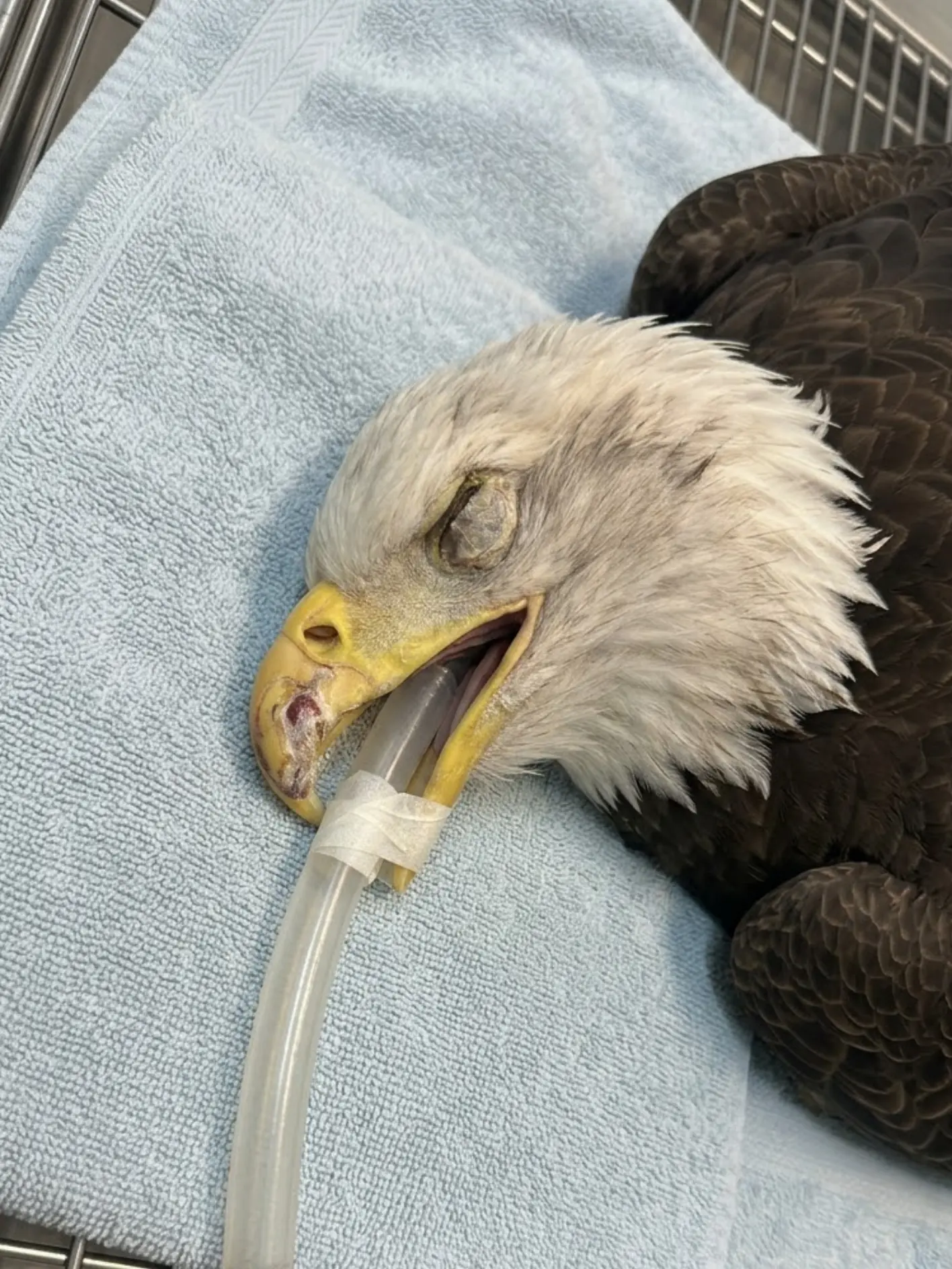
At first glance, it did not immediately look like an emergency.
Raptors are not particularly tidy eaters. Pieces of meat, blood, and other food material can occasionally remain around the beak after feeding. Seeing something dried along the edge of a Bald Eagle’s bill is therefore not automatically unusual.
But this eagle has been in long-term care, and our staff know the normal appearance of its beak very well.
Something looked different.
The material was in an unusual location, and the contour of the left side of the beak did not look the way it normally did.
Rather than assume it was leftover food, staff brought the eagle to the hospital for a closer examination.
Once the area was cleaned, the extent of the problem became clear.
A substantial portion of the keratin covering the left side of the beak had broken away. The damage extended along the side and toward the tip, leaving portions of the underlying structure exposed down to the bone.
What had appeared from a distance to be dried food was actually hiding a significant beak injury.
The exact cause remains unknown.
For now, the medical team has cleaned and disinfected the damaged area, placed protective bone cement over the exposed portion, and started the eagle on antibiotics and pain medication. The injury will be reassessed after approximately one week, but the longer-term prognosis for the beak itself remains uncertain.
The greatest immediate concern is infection.
The larger question may take much longer to answer:
Will enough healthy keratin grow back to allow this eagle to continue eating, preening, and living comfortably with an already abnormal beak?
The First Sign Was Easy to Mistake for Food
The injury was discovered because someone familiar with the eagle noticed a subtle change.
There was dried blood near the left side of the beak and material that initially resembled meat residue.
That interpretation was reasonable.
A Bald Eagle uses its beak to tear food into manageable pieces, and small amounts of blood or tissue can sometimes remain on the outer surface after a meal.
But the appearance was unusual for this particular bird.
The left side looked irregular.
The dried material was not sitting on the surface in the way ordinary food residue usually does.
Closer inspection was warranted.
That decision illustrates an important part of caring for permanent education animals: familiarity with an individual bird makes small changes easier to recognize.
A veterinary examination may begin because of something dramatic — a bird suddenly unable to stand, an obvious fracture, or active bleeding.
Other times it begins because a staff member notices that something simply does not look normal.
In this case, that small observation led to the discovery of a much deeper injury.
The Protective Keratin Layer Had Broken Away
After the eagle was safely restrained and the dried material was removed, veterinary staff could clearly see that a significant portion of the outer beak had been lost.
The damage affected the left side of the beak and extended toward the tip.
Much of the protective keratin layer was missing.
In several places, the injury reached the underlying bony structure.
That distinction is important.
A minor chip involving only the superficial edge of a beak may require monitoring but can sometimes be relatively straightforward.
Loss of the keratin covering down to exposed bone is a different situation.
The outer beak is not merely a decorative shell.
It protects sensitive living structures beneath it and provides the hard working surface a raptor uses every day.
Once that protection is lost, the deeper tissues become vulnerable to contamination, pain, inflammation, and infection.
A Raptor’s Beak Is More Complex Than It Looks
The visible outer portion of a bird’s beak is composed largely of keratin, the same broad class of structural protein found in feathers and claws.
In birds, the keratinized outer covering of the beak is known as the rhamphotheca.
Beneath that protective layer are living tissues and bone.
The keratin covering grows continuously and is shaped through normal wear.
For a Bald Eagle, the beak has to withstand considerable mechanical stress.
It is used to:
- tear flesh,
- manipulate food,
- assist with preening,
- move objects,
- and perform normal daily behaviors.
The hooked tip provides leverage when the bird pulls pieces of meat away from food being held with the feet.
A defect that alters the shape, strength, or growth of the beak can therefore affect far more than appearance.
This eagle was already starting from an unusual anatomical situation.
This Eagle Has Had a Lifelong Beak Abnormality
The bird’s beak was not completely normal before this new injury occurred.
When the eagle was younger, it suffered a severe avian pox lesion that affected the developing beak.
Although the active disease resolved long ago, the lesion left a permanent deformity.
As a result, the beak has had an abnormal structure for much of the eagle’s life.
That history matters because tissue that develops abnormally may not distribute mechanical stress in the same way as a normal beak.
Veterinarians believe the pre-existing deformity may have created an area of structural weakness.
However, that remains a possibility rather than a confirmed explanation.
The team cannot say that the old pox lesion directly caused the new break.
It can only say that the eagle already had a compromised beak before this episode.
The Exact Cause of the New Injury Is Unknown
No one witnessed the moment the beak was damaged.
There was no known collision.
No observed fall.
No documented aggressive encounter.
No specific object was identified that could explain the missing section.
Based on when the injury was discovered and the bird’s recent routine, staff believe it may have occurred on Sunday.
That was a relatively quiet day at the center.
Only essential veterinary personnel and a reception coordinator were working, and the eagle was not scheduled to receive food that day.
That information helps narrow the likely time window.
It does not establish a mechanism.
The fact that the eagle was not fed that day may make an ordinary feeding-related accident less obvious as an explanation, but there is still insufficient evidence to determine what happened.
The medical team is therefore treating the injury rather than trying to force certainty onto an event no one saw.
The Wound Was Cleaned Before Anything Else
Once the full extent of the damage could be seen, the exposed region was carefully cleaned.
Dried blood, food contamination, and debris were removed.
The area was disinfected so veterinary staff could assess the remaining keratin and the exposed underlying structures.
Cleaning served two purposes.
First, it allowed clinicians to determine how deep the injury actually extended.
Second, it reduced the amount of material that could potentially contribute to bacterial contamination.
The beak is constantly exposed to microorganisms.
A raptor uses it during feeding.
It contacts meat.
It contacts feathers.
It is involved in preening.
A deep defect with exposed bone therefore requires much closer infection monitoring than an uncomplicated superficial chip.
Bone Cement Was Used as a Temporary Protective Barrier
After the wound was cleaned and evaluated, the veterinary team applied bone cement over the exposed area.
This should not be confused with completely reconstructing the eagle’s beak.
The material is serving primarily as protection.
The natural keratin normally shields the underlying structures from direct exposure.
With a large portion missing, the bone cement can provide a temporary barrier while clinicians observe how the injury heals.
Its purpose is to help protect the exposed region from:
- contamination,
- repeated mechanical irritation,
- food particles,
- and direct contact.
The material also gives the injured area some temporary protection while the team waits to determine whether natural keratin growth will begin restoring the missing surface.
It is not considered a substitute for a healthy natural beak.
Infection Is the Immediate Medical Concern
The deepest portion of the injury reaches the bone.
That creates a significant concern for infection.
Superficial skin and keratin injuries may often be managed locally.
Once bone is exposed, bacteria have a potential route into deeper structures.
One complication veterinary staff will be watching closely for is osteomyelitis, an infection involving bone.
The eagle has not been diagnosed with osteomyelitis.
The team’s goal is to prevent it.
During the coming days, staff will monitor for changes such as:
- increased swelling,
- discharge,
- odor,
- tissue discoloration,
- worsening pain,
- changes in appetite,
- or deterioration around the protected site.
Any of those findings could indicate that the injury is not progressing normally.
Antibiotics Were Started Because of the Risk
Given the depth of the defect and the exposed bone, the eagle is being treated with antibiotics under veterinary supervision.
The medication is intended to address the risk of bacterial contamination associated with the injury.
As with all wildlife patients, the treatment plan will be adjusted according to clinical response.
The medical team will not rely on antibiotics alone.
The condition of the wound itself remains equally important.
Cleaning.
Protection.
Monitoring.
Nutrition.
Pain control.
And time.
All of those factors will influence the outcome.
The Injury Is Also Painful
Keratin itself is not the same as living soft tissue, but the structures beneath it are sensitive.
A defect deep enough to expose underlying bone can cause considerable discomfort.
The eagle is therefore also receiving pain medication.
Pain control is important for reasons beyond comfort.
An eagle experiencing significant beak pain may stop eating.
It may avoid preening.
It may repeatedly rub the injured area.
Stress can increase.
Those behaviors can complicate healing.
Appropriate analgesia helps reduce those secondary problems while the injured region is stabilized.
Feeding Will Be Watched Closely
One of the most important questions over the next several days will be how well the eagle can eat.
Bald Eagles normally use their feet and beak together.
The feet hold the food.
The curved beak tears pieces away.
A weakened or painful beak can make that process difficult.
For the time being, staff can modify food presentation if necessary.
Meat can be prepared into pieces that require less force to tear.
The bird’s appetite can be monitored independently of its ability to process larger food items.
What staff do not want to do is deliberately place unnecessary mechanical stress on the injured beak simply to see whether it can tolerate it.
Healing comes first.
The Beak’s Function Extends Beyond Feeding
Food handling is only one concern.
The beak is also essential to feather maintenance.
Bald Eagles spend considerable time preening.
During preening, the bird uses the beak to clean and realign feathers.
This matters for insulation, waterproofing, and flight.
A permanent beak defect can therefore create secondary problems even in a bird that receives food in human care.
The eagle must still be able to maintain its plumage and perform normal behaviors.
That is why veterinarians are thinking not only about wound closure, but also about long-term function and quality of life.