The young Bald Eagle arrived at Northwoods Wildlife Rehabilitation Center on Tuesday afternoon carrying two problems that would each have been serious on their own.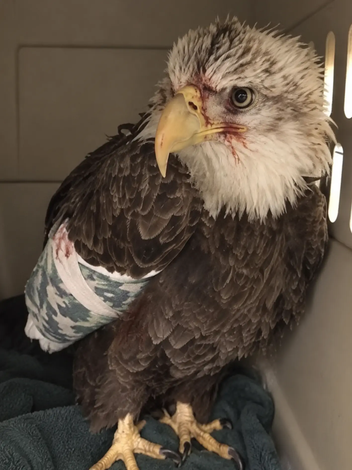
One could be seen on an X-ray.
The other could only be found in the blood.
The eagle had been recovered earlier that day from a rain-soaked field in Eau Claire County, Wisconsin, after a concerned landowner reported a large raptor on the ground that appeared unable to fly.
Volunteer wildlife transporter Rachel Donovan responded to the call.
What followed was considerably more difficult than the eagle’s condition initially suggested.
Although the bird could not take flight and one wing was being held abnormally, it still had enough strength to move quickly through the wet grass whenever Donovan approached.
Rain had already softened the field.
Every step became more difficult as mud collected around her boots.
The eagle alternated between short runs, awkward hops, and attempts to create distance while keeping the injured wing close to its body.
For several minutes, the severely compromised bird made its rescuer work for every foot of ground she gained.
Eventually, Donovan was able to contain it safely.
Only later would the veterinary team discover just how depleted the eagle actually was.
Its body condition score was 1 out of 5.
Its upper wing was fractured.
Follow-up radiographs would show that the break was more complicated than first appreciated.
And blood testing would reveal another significant concern:
lead exposure.
“A heartfelt thank-you goes to Rachel,” the rehabilitation team said. “This bird was extremely thin and had a broken wing, but it still sent her across a muddy field in heavy rain before she could safely contain it.”
That resistance was one of the few encouraging signs in the eagle’s first hours of care.
Its body was severely depleted.
Its behavior showed that some fight remained.
The Eagle Was Grounded but Still Defensive
When Donovan first reached the field, the young eagle was clearly abnormal.
A healthy Bald Eagle with open space around it would normally attempt to leave before allowing a person to approach closely.
This bird could not.
One wing remained lower and closer to the body than the other.
It occasionally opened the healthy wing and made short evasive movements, but sustained flight was impossible.
The bird remained alert.
Its eyes followed Donovan.
Its feet came forward defensively when she moved within reach.
That reaction mattered.
A severely weakened eagle can still cause serious injury with its talons.
Donovan therefore avoided forcing the bird into repeated escape attempts and concentrated on safely containing it for transport.
Once secured, the eagle was placed in a padded carrier and driven to Northwoods Wildlife Rehabilitation Center.
The receiving team prepared for trauma.
What they found on intake suggested that the bird had been struggling for much longer than the few minutes witnessed in the field.
A Body Condition Score of 1/5 Changed the Medical Picture
The eagle was profoundly thin.
On physical examination, the keel of the sternum was sharply prominent beneath the breast.
The pectoral muscles — the large muscles responsible for powering flight — were severely reduced.
Very little fat reserve could be identified.
The veterinary team assigned the bird a body condition score of 1/5, indicating severe loss of muscle and stored energy.
This was not simply a naturally lean wild bird.
It was a patient with minimal physiologic reserve.
For Dr. Emily Carter, the center’s attending veterinarian, that finding immediately complicated the case.
“A wing fracture is already a major injury in an eagle,” Carter said. “But when the bird has almost no body reserves left, you have to think about much more than the bone. You have to ask whether the patient can tolerate anesthesia, whether it can heal efficiently, and whether there is another underlying problem contributing to that condition.”
The eagle was also dehydrated.
Its feathers were disordered.
There were minor abrasions around the head and body consistent with time spent grounded and struggling.
But the most obvious orthopedic problem involved the injured wing.
The First X-Rays Confirmed a Humeral Fracture
Palpation of the affected wing produced pain and abnormal movement in the upper portion of the limb.
Radiographs confirmed a fracture of the humerus, the large bone extending between the shoulder and elbow.
In a Bald Eagle, this is not a minor skeletal injury.
The humerus forms a major structural component of the wing and lies within a region subjected to significant forces during powered flight.
A fracture that heals in poor alignment may alter the geometry of the entire wing.
Even small losses in motion or symmetry can matter when an eagle must generate lift, climb, turn, brake, and land.
The initial films showed that the fracture was displaced.
The wing was stabilized against the body while the veterinary team addressed the bird’s immediate medical needs.
Pain management was provided as clinically appropriate.
Fluids were administered to correct dehydration.
The eagle was placed in a quiet enclosure with restricted movement.
At this stage, the wrap around the injured wing was not considered definitive treatment.
It was a temporary measure intended to reduce motion and pain while the team gathered more information.
“The wrap gives us time,” Carter explained. “It doesn’t solve a humeral fracture.”
Follow-Up Radiographs Changed the Conversation
Once the eagle was more stable and additional diagnostic positioning became possible, the team obtained follow-up radiographs.
Those images revealed that the fracture was more serious than the initial films had suggested.
The break showed greater displacement when viewed from additional angles.
There also appeared to be more complexity around the fracture site than had been obvious on the first examination.
The question was no longer simply how long the wing should remain stabilized.
The new question was whether the bone could be surgically reconstructed with a realistic expectation of restoring flight.
For a wildlife patient, that distinction is critical.
A fracture can heal anatomically without producing a releasable animal.
For a Bald Eagle, the standard is not simply union of the bone.
The wing must eventually function with enough precision to support normal survival in the wild.
“The second set of images made us much more cautious,” Carter said. “We had to determine whether this was a fracture that could be repaired well enough for flight, not just whether we could make the bone heal.”
The radiographs were forwarded to Dr. Michael Reeves, an avian orthopedic consultant, for review.
The veterinary team wanted an assessment of the fracture geometry, degree of displacement, proximity to important joints, and likelihood that surgical fixation could restore appropriate alignment.
But before any surgical plan could be finalized, another test result complicated the case further.
Blood Testing Detected Lead
Because the eagle was so severely underweight, the veterinary team performed a broader diagnostic evaluation.
Grounded Bald Eagles can be presented with trauma, starvation, infectious disease, toxic exposure, neurologic illness, or several problems occurring simultaneously.
Lead exposure is an important consideration in scavenging raptors.
The eagle’s blood was therefore tested.
The result showed an elevated lead concentration significant enough to require clinical attention.
The discovery did not explain the broken humerus.
There was no evidence that lead itself had directly fractured the bone.
But it could help explain why the eagle had arrived with so little reserve.
Lead toxicosis in birds can be associated with weakness, reduced appetite, gastrointestinal dysfunction, neurologic abnormalities, impaired coordination, and progressive loss of condition.
An affected eagle may become less capable of feeding successfully.
It may also become more vulnerable to secondary trauma.
In this case, however, the rehabilitation team could not establish exactly how the eagle had encountered the metal.
That distinction was important.
The Source of the Lead Was Unknown
Bald Eagles are opportunistic scavengers.
They may consume carcasses or discarded animal remains in addition to catching live prey.
One recognized route of lead exposure in raptors is ingestion of small ammunition fragments left in animal tissue.
Fragments can remain in carcasses or gut piles and may be swallowed by scavenging birds.
Once inside the gastrointestinal tract, lead can be absorbed into the bloodstream.
But no one had observed this particular eagle feeding on a contaminated carcass.
No source could be confirmed.
The team therefore documented lead exposure without claiming a specific origin.
“The blood tells us the eagle has been exposed,” Carter said. “It does not tell us precisely where that exposure occurred.”
That cautious distinction allowed the medical team to address the toxicologic problem without turning an unknown exposure history into an unsupported conclusion.
One Injury Was Visible on X-Ray; the Other Was Visible in the Blood
The two diagnoses created a difficult interaction.
The fractured humerus required stability and potentially surgery.
The lead exposure required systemic treatment.
The severe emaciation meant the bird had very limited metabolic reserves for either problem.
A strong, well-conditioned eagle with an otherwise isolated fracture may tolerate anesthesia differently from a bird at a body condition score of 1/5.
Anesthesia places demands on the cardiovascular and respiratory systems.
Surgery adds physiologic stress.
Healing requires protein, energy, oxygen delivery, and adequate circulation.
A severely depleted animal may have less capacity to tolerate each stage.
That meant the team could not simply see a repairable fracture and proceed immediately.
Repairing the bone would mean little if the patient was not strong enough to survive the repair.
Stabilization Became the First Priority
For the first phase of care, the goal was to improve the eagle’s general condition while preventing further damage to the wing.
The fracture remained immobilized.
The enclosure was designed to prevent unnecessary movement.
No high perch was offered.
The bird needed to conserve energy.
Hydration was corrected gradually.
Pain was controlled.
Bloodwork was reviewed for evidence of additional abnormalities.
The medical team monitored packed cell volume and other parameters relevant to the bird’s ability to tolerate continued treatment.
Lead management was initiated as clinically indicated.
Where chelation therapy is appropriate, medications are used under veterinary supervision to bind lead and assist its removal from the body.
The eagle would require repeated assessment to determine whether blood lead levels were responding.
Nutrition presented another challenge.
Feeding an Eagle at 1/5 Body Condition Requires More Than Simply Giving It a Large Meal
It might seem intuitive that a severely underweight animal should be given as much food as possible immediately.
Clinical nutritional rehabilitation can be more complicated.
A body that has been underfed for an extended period may not respond normally to sudden high caloric intake.
The gastrointestinal system needs to function.
Hydration must be adequate.
Electrolyte and metabolic balance matter.
The eagle therefore received a carefully managed nutritional plan.
Small meals were introduced and monitored.
The team watched whether the bird swallowed normally.
Whether food remained down.
Whether fecal output appeared appropriate.
Whether appetite increased.
The objective was not simply to add weight to the scale as rapidly as possible.
It was to rebuild physiologic stability safely.
“The first goal is not to make the bird look heavier,” Carter said. “The first goal is to make sure the body can use what we are giving it.”
The Eagle Remained Surprisingly Defensive
Despite its condition, the bird continued behaving like a wild eagle.
During necessary handling, it watched staff closely.
Its feet remained dangerous.
The beak opened defensively.
It attempted to create distance whenever possible.
Those reactions made examinations more difficult.
They were also encouraging.
The veterinary team had a patient that was profoundly thin but still neurologically engaged with its surroundings.
“The body condition told us how depleted the eagle was,” one rehabilitator said. “Its attitude told us it had not stopped fighting.”
That attitude alone could not determine prognosis.
But it mattered.
A bird becoming progressively less responsive despite stabilization would have been much more concerning.
Instead, the eagle remained alert.
The First Meals Were Small but Important
The bird began eating.
Not large amounts at first.
But enough to demonstrate that appetite and gastrointestinal function were present.
Over the following days, interest in food increased.
Each meal supplied energy that the bird desperately needed.
Staff recorded weight carefully.
One measurement meant little.
The trend mattered.
Was the eagle maintaining weight?
Then gaining?
Was muscle loss continuing?
Was hydration improving?
The same principle applied to blood testing.
The veterinary team needed trends, not isolated numbers.
Lead concentration would be reassessed.
Other blood values would be followed.
The goal was to build enough evidence that the bird was moving toward greater stability rather than simply appearing more alert.
The Humeral Fracture Could Not Wait Indefinitely
While the team worked on nutrition and lead exposure, another clock was running.
Fractures begin healing whether or not the final treatment plan has been decided.
Callus forms.
Soft tissues contract.
Bone fragments may begin consolidating where they sit.
That can be beneficial when alignment is appropriate.
It can make later reconstruction more difficult when alignment is poor.
This created a difficult balance.
The eagle needed stabilization before anesthesia.
The wing could not remain in an unfavorable position indefinitely.
The orthopedic consultation therefore became urgent.
The radiographs had been sent to Reeves along with details from the physical examination.
He reviewed the configuration of the break.
The amount of displacement.
The apparent condition of the surrounding bone.
Its position relative to the elbow and shoulder.
And the likely mechanical consequences if it healed without correction.
His assessment would help determine whether surgery offered a meaningful chance of returning the eagle to flight.
“Surgically Repairable” and “Releasable” Are Not the Same Thing
Wildlife orthopedic decisions differ in one important respect from many companion-animal cases.
The final standard is independence.
A procedure might successfully stabilize a fracture.
The bird might live comfortably afterward.
But if normal flight cannot reasonably be restored, the outcome may still be inadequate for release.
A Bald Eagle must generate powerful symmetrical wingbeats.
The shoulder and elbow need sufficient range of motion.
Wing length and alignment must remain compatible with efficient aerodynamics.
The bird has to sustain flight, not simply flap across a room.
That means veterinarians must consider both the technical repair and the likely functional result.
Reeves considered the fracture potentially reconstructable, but the prognosis for eventual unrestricted flight remained guarded.
The bird’s poor systemic condition made the decision even more cautious.
The bone might be repairable.
The patient still had to become a safer anesthetic candidate.
Lead Exposure Complicated the Timing
Lead treatment and orthopedic treatment could not be considered completely separately.