An adult bald eagle found grounded in Kitsap County, Washington, was brought to a licensed wildlife rehabilitation and raptor care center after county animal services officers discovered the bird unable to fly and breathing with obvious difficulty.
At first, the case appeared to involve trauma.
The eagle had abrasions on its legs and feet. There was bruising over portions of the pectoral musculature. Its posture was abnormal, and the bird appeared weak enough that responders were able to contain it safely for transport.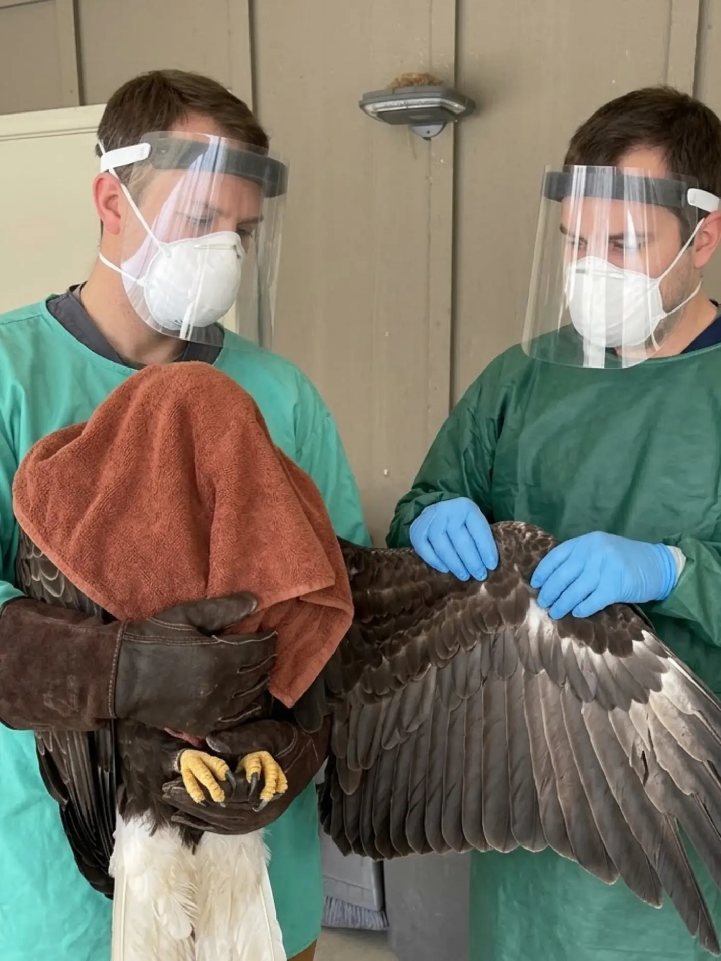
But once the eagle reached the rehabilitation center, veterinary staff began seeing signs that the visible injuries did not fully explain what was happening.
The bird repeatedly shook and nodded its head in an abnormal pattern.
Its coordination was impaired.
It continued to breathe heavily, matching the respiratory distress described by the people who first found it.
Within hours, the neurological signs became more pronounced.
Several explanations remained possible at intake. Direct trauma could produce neurological abnormalities. Toxic exposure, including certain rodenticides or other environmental contaminants, could cause weakness and abnormal movements. A systemic infectious disease could also produce neurological and respiratory signs and might even explain how the bird became injured in the first place.
Because of that uncertainty, the eagle was treated as more than a routine trauma patient.
Staff used enhanced personal protective equipment during examination, including protective gowns, gloves, masks and face shields. The bird was isolated from other avian patients. Diagnostic samples were collected, including material submitted for avian influenza testing.
The abrasions and bruising were treated.
Supportive care was initiated.
But despite those measures, the eagle deteriorated rapidly.
The neurological signs worsened during the first day of hospitalization, and the bird died within 24 hours of admission.
The laboratory result had not yet returned.
It arrived afterward.
The eagle tested positive for highly pathogenic avian influenza, or HPAI.
The result did not prove that every abrasion or area of bruising had been caused by the infection. But it provided an important explanation for the rapid neurological decline, respiratory abnormalities and profound loss of normal function observed after admission.
“The traumatic injuries were real, but they did not fully explain what we were seeing neurologically,” a veterinarian involved in the case said. “The repeated head movements, inability to fly normally and respiratory distress meant we had to consider a systemic or infectious process from the beginning.”
The Eagle Was Found Grounded in Kitsap County
Bald eagles are a familiar presence around Kitsap County and the wider Puget Sound region.
The landscape provides extensive shoreline habitat, wetlands, forested areas and open water where eagles can feed on fish, waterfowl, carrion and other available food sources.
Seeing an adult eagle perched near the water or crossing overhead is not unusual.
Seeing one remain on the ground and fail to fly when approached is different.
County animal services officers were called after the bird was reported in a compromised condition.
When responders arrived, they observed that the eagle was unable to take off normally.
The bird also appeared to be breathing harder than expected.
That combination immediately raised concern.
A grounded eagle may have a broken wing.
It may have head trauma.
It may be intoxicated.
It may be severely debilitated by infection.
Or it may be experiencing more than one problem at the same time.
The officers approached cautiously.
Even an impaired bald eagle remains capable of inflicting serious injury with its feet and talons.
The bird’s legs and feet had to be controlled while the wings were kept safely against the body.
Once contained, the eagle was placed into a ventilated transport carrier and moved to the rehabilitation center for evaluation.
The First Examination Revealed Real Trauma
The veterinary team began with a full physical examination.
There were abrasions involving the legs and feet.
Bruising was visible or palpable over portions of the pectoral musculature.
The feathers in several areas were disturbed, consistent with the bird having been on the ground or having experienced some form of physical impact.
Those findings initially kept trauma high on the list of possibilities.
A bird that strikes a structure or falls awkwardly can sustain bruising without breaking a bone.
A head impact can also cause neurological signs.
But the pattern did not fit neatly.
The eagle was showing more than pain or weakness.
Its head movements were abnormal.
The bird repeatedly shook or nodded the head in ways that did not appear voluntary.
Its coordination was impaired.
The respiratory effort persisted.
Those signs pushed the examination beyond the question of “Where is the injury?”
The more important question became:
Why is the nervous system behaving this way?
The Respiratory Distress Could Not Be Ignored
The people who initially found the eagle had described it as breathing hard.
That observation remained relevant after admission.
The bird continued to show increased respiratory effort.
At times the beak was held open.
The pattern was not easily explained by the superficial abrasions alone.
Pain and stress can increase respiratory rate in wildlife.
Handling itself can temporarily make a bird breathe faster.
But persistent respiratory difficulty, combined with abnormal neurological signs, raises concern for a broader systemic process.
That is when the differential diagnosis had to expand.
Neurological Signs Changed the Entire Case
The repetitive head movements became one of the most important observations.
A bald eagle that is simply weak from orthopedic trauma may hold a wing abnormally or refuse to stand.
A bird with neurological disease may:
- lose coordination,
- show abnormal head movements,
- struggle to maintain posture,
- become disoriented,
- or lose the ability to fly despite having intact wings.
The eagle in this case showed enough neurological abnormality that the veterinary team could not treat the situation as straightforward trauma.
There were several plausible explanations.
Head injury remained possible.
Toxin exposure remained possible.
Infectious disease became increasingly important.
No single explanation could yet be confirmed.
That uncertainty dictated how the bird had to be managed.
Trauma Was Still a Legitimate Possibility
The abrasions and pectoral bruising mattered.
The eagle may have struck the ground.
It may have collided with an object.
It may have fallen after becoming weak.
The bird may also have suffered trauma before any neurological illness developed.
At intake, veterinarians could not know which event came first.
That distinction matters.
If trauma occurred first, neurological abnormalities might have been secondary to head injury.
If systemic disease occurred first, the eagle may have lost coordination in flight and crashed, producing the abrasions and bruising afterward.
Both sequences were medically plausible.
Only testing and the clinical course could help clarify the situation.
Toxic Exposure Was Also Considered
Wildlife clinicians evaluating a neurologically abnormal raptor must consider toxins.
Some rodenticides can cause systemic illness through different mechanisms.
Other environmental contaminants can affect neurological function.
Lead exposure can also produce weakness and neurological abnormalities in bald eagles, particularly because the species may scavenge animal remains.
The rehabilitation team therefore had good reason not to assume the bird’s symptoms came from one cause.
A poisoned eagle may fall.
A neurologically impaired eagle may injure itself.
Trauma and toxin exposure can exist together.
The same is true of infectious disease.
The case had to remain open diagnostically.
Infectious Disease Became a Serious Concern Early
The combination of neurological signs and respiratory abnormalities meant an infectious process could not be ruled out.
That possibility had immediate implications beyond treatment.
A suspected infectious disease in a wild bird creates a biosecurity issue.
Staff have to think not only about the patient, but also about:
- other birds in the facility,
- contaminated equipment,
- clothing,
- surfaces,
- transport materials,
- and potential exposure of personnel.
The eagle was therefore handled with enhanced precautions while testing was pending.
The Protective Equipment Seen During Examination Had a Purpose
During the initial examination, staff wore protective gowns, gloves, respiratory protection and face shields while one handler controlled the bird’s feet and body and another examined the wing and soft tissues.
A towel was used over the head during portions of restraint.
That serves several purposes.
Reducing visual stimulation can decrease stress.
It helps limit sudden defensive movement.
It also makes controlled examination safer for both the bird and staff.
The protective equipment was not proof that HPAI had already been diagnosed.
At that stage, the team did not yet know.
The equipment reflected the fact that an infectious disease could not safely be excluded.
The Eagle Was Isolated From Other Avian Patients
Once infectious disease entered the differential diagnosis, keeping the bird separated from other birds became essential.
A rehabilitation center may house multiple raptors, waterbirds or other wildlife patients at the same time.
A pathogen capable of spreading among birds can create consequences far beyond one individual case.
The eagle was therefore placed in an isolation area with restricted access.
Equipment used for the bird could be kept dedicated or managed separately.
Staff contact was minimized.
Handling occurred only when medically necessary.
This approach simultaneously reduced disease-transmission risk and lowered stress on a critically ill eagle.
The Physical Injuries Were Still Treated
Isolation did not mean the traumatic injuries were ignored.
The abrasions on the legs and feet were examined and cleaned.
Bruised tissues were evaluated.
The wings were checked carefully for range of motion, swelling and evidence of more serious structural injury.
Pain management could be provided when clinically appropriate.
The bird’s hydration and overall condition were assessed.
The team was effectively managing two possibilities at once.
A trauma case.
And a possible systemic infectious case.
That is often necessary in wildlife medicine before diagnostic results return.
Imaging Did Not Explain the Severity of the Signs
If the eagle was stable enough for imaging, radiographs could help determine whether a major fracture or obvious skeletal injury explained the inability to fly.
The critical point in this case was that no large displaced fracture emerged that could account for the rapid neurological deterioration.
The bird had trauma.
But the skeletal findings did not explain why the nervous system was progressively failing.
That discrepancy became increasingly important.
If the physical injury remained relatively stable while neurological function deteriorated hour by hour, the clinicians had to look elsewhere for the primary process.
Samples Were Submitted While Supportive Care Began
Diagnostic testing was initiated.
Samples were collected for avian influenza testing along with other laboratory work considered appropriate to the case.
Those tests required time.
The bird did not have the luxury of waiting untreated.
Supportive care began while the laboratory work was pending.
That could include fluid support, careful temperature management, quiet isolation and respiratory support where available and indicated.
Because neurological patients may have impaired swallowing, feeding decisions had to be made cautiously.
A bird that cannot protect its airway normally is at risk of aspiration if food or fluid is administered incorrectly.
In a critically ill wildlife patient, sometimes the safest intervention is to minimize handling until the bird can tolerate more.
HPAI Was Not Diagnosed at the Door
This point is important.
The rehabilitation team did not look at the eagle and immediately know it had highly pathogenic avian influenza.
The clinical signs overlap with many other conditions.
A grounded bald eagle with head movements and weakness could have:
- trauma,
- poisoning,
- neurological disease,
- metabolic abnormalities,
- or infection.
That is why laboratory confirmation matters.
The correct approach is not to guess.
It is to isolate appropriately, stabilize when possible, collect diagnostic samples and follow the clinical evidence.
The Neurological Signs Worsened Rapidly
During the hours after admission, the eagle’s condition deteriorated.
The abnormal head movements became more pronounced.
The bird had increasing difficulty maintaining normal posture and coordination.
Its respiratory effort remained concerning.
It became progressively less capable of functioning normally.
This rate of decline stood out.
The abrasions themselves were not suddenly becoming catastrophic.
The bruising had not changed enough to explain the deterioration.
Something systemic was progressing faster than the visible injuries.
That realization further strengthened concern for an underlying neurological or infectious disease.
The Bird Died Before the Test Result Returned
Despite supportive care, the eagle did not stabilize.
Its neurological condition continued to worsen.
The bird died within the first 24 hours after admission.
At that point, the rehabilitation team still did not have the final avian influenza result.
That is an important detail because it reflects the reality of wildlife infectious-disease cases.
Diagnostic laboratories do not always return answers before a critically ill animal reaches the end of its clinical course.
The treatment team may be forced to make biosecurity and supportive-care decisions under uncertainty.
In this case, that uncertainty lasted longer than the eagle survived.
The HPAI Result Arrived Afterward
The laboratory result later confirmed that the bald eagle was positive for highly pathogenic avian influenza.
That finding changed the interpretation of the entire case.
The respiratory abnormalities now fit a known infectious process.
So did the neurological signs.
The rapid deterioration also became more understandable.
The result did not prove exactly when the eagle became infected.
It did not reveal where exposure occurred.
And it did not establish whether every physical injury was secondary to infection.
But it provided a strong explanation for the systemic disease veterinarians had been witnessing.
“Unfortunately, the neurological decline was extremely rapid,” the veterinarian said. “The eagle died before the avian influenza result was returned, and that positive result later provided an important explanation for the progression we had observed.”
HPAI Can Present With Neurological Signs
Highly pathogenic avian influenza is often discussed in relation to poultry or waterfowl, but wild raptors can also be affected.
Clinical presentation varies.
Some infected birds may show weakness.
Others develop neurological abnormalities.
Loss of coordination can occur.
Abnormal head movements may appear.
Some birds develop respiratory signs.
Others may deteriorate rapidly with relatively little warning.
In raptors, scavenging behavior may create opportunities for exposure to infected birds or carcasses.
But the exact route of exposure for this eagle was not identified.
That uncertainty should remain explicit.