An adult bald eagle brought to a small wildlife stabilization clinic in Chatham County, Georgia, arrived in a condition that immediately alarmed the rehabilitation staff.
The bird was lying on its back.
It could not turn itself upright.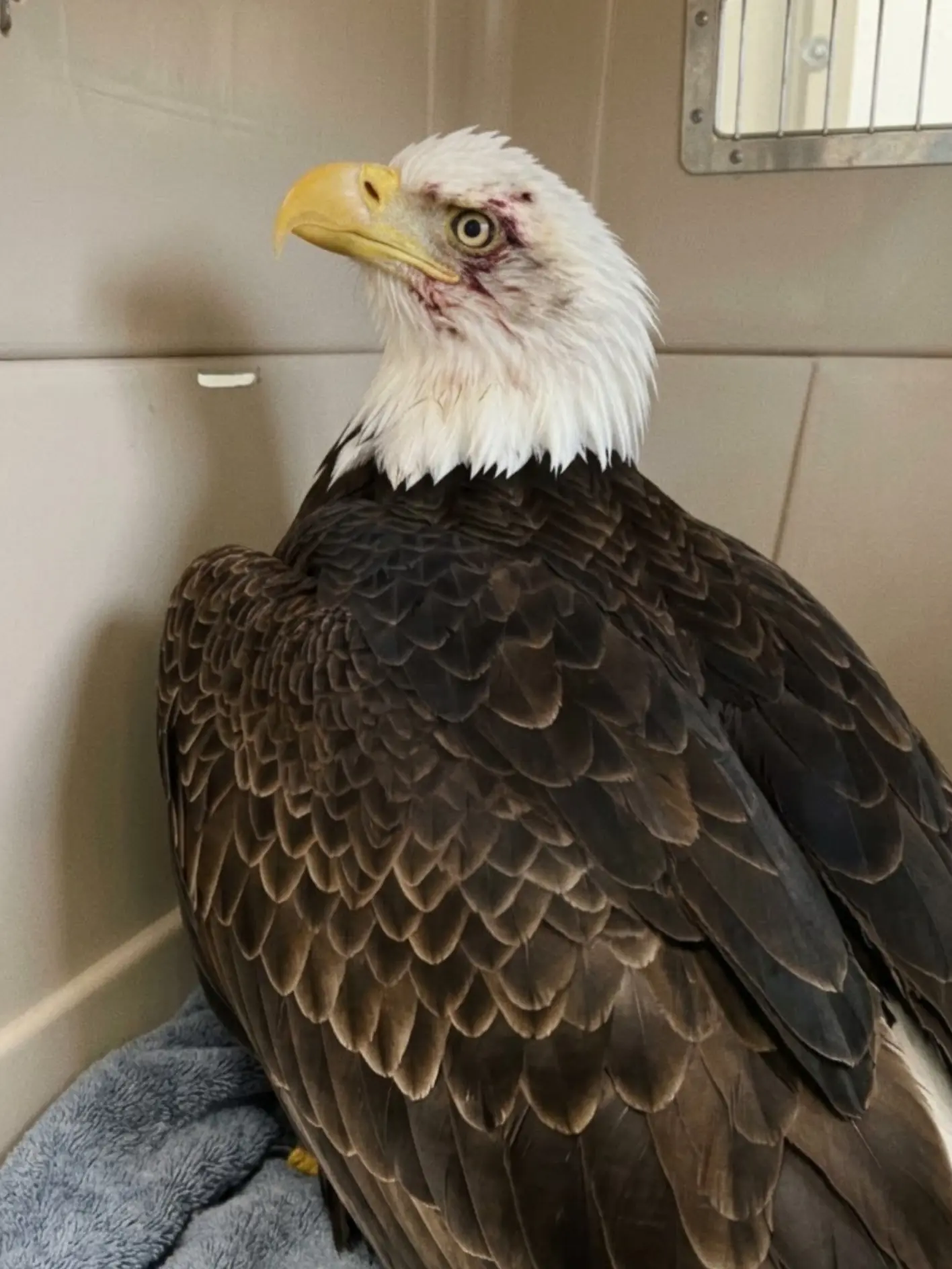
Its breathing was rapid and labored, and repeated seizure activity caused its body to stiffen and contract without warning. The eagle showed poor control of its head and limbs, and its pupils did not appear to respond equally. Even when the seizures temporarily stopped, it had difficulty focusing visually on the people and movement around it.
There was no time to wait for a definitive toxicology result.
The clinical picture raised immediate concern for a severe neurological emergency, with toxic exposure high on the list of possible causes.
Staff began supportive treatment almost immediately.
Subcutaneous fluids were administered to address dehydration and support circulation. Activated charcoal was carefully introduced into the gastrointestinal tract in an effort to bind certain toxins that might still be present and reduce additional absorption. The eagle was then placed in a towel-lined carrier that supported its body and reduced the risk of further injury if another seizure occurred.
The carrier was moved into a quiet, dark, warm room.
Light was reduced.
Noise was minimized.
Handling was kept to an absolute minimum.
At that point, no one at the clinic could say with confidence whether the eagle would survive the night.
By the following morning, however, something had changed.
The bird was more alert.
The seizures had become far less frequent.
Its breathing was somewhat steadier.
But the improvement was incomplete.
The eagle still could not stand normally. Its balance remained poor, and it continued to have difficulty focusing its gaze.
The staff repeated supportive fluids, administered additional activated charcoal when medically appropriate, and began providing a highly digestible nutritional formula formulated for carnivorous wildlife.
The bird had survived the first critical phase.
The larger question was whether its nervous system could recover.
The Eagle Arrived in a Full Neurological Crisis
Wildlife patients often arrive without a known history.
There may be no witness to the event that injured or sickened them.
There is no owner who can say when symptoms began.
And there is no way for the animal to communicate what it was doing before it became incapacitated.
Veterinary staff therefore have to work backward from the clinical signs.
In this case, those signs were severe.
The eagle’s inability to right itself was one of the most alarming.
A healthy bird placed on its back will instinctively attempt to roll or push itself into a normal upright position.
This eagle could not.
That loss of postural control suggested significant neurological dysfunction, profound weakness, or both.
Repeated seizures added another major concern.
The episodes were not simply tremors or brief moments of instability. Staff observed recurrent involuntary contractions consistent with severe neurological disturbance.
The abnormal pupils were another clue.
One appeared more dilated than the other, and the bird struggled to maintain normal visual focus.
Taken together, the signs suggested that the problem extended well beyond ordinary exhaustion or a localized wing injury.
Something was affecting the bird’s nervous system.
Toxic Exposure Became One of the Leading Concerns
Several conditions can produce seizures and loss of coordination in a bald eagle.
Head trauma is one.
Severe metabolic disease is another.
Some infections can affect the nervous system.
Toxins can as well.
Because the eagle showed multiple neurological abnormalities without an immediately obvious external injury severe enough to explain all of them, staff considered poisoning an important possibility from the beginning.
That did not mean they knew what toxin was involved.
They did not.
The appropriate description was suspected toxic exposure, not confirmed poisoning from a specific substance.
Blood testing and additional diagnostic work would be needed to move from suspicion to diagnosis.
But emergency wildlife medicine often requires clinicians to begin supportive treatment before every laboratory answer is available.
A severely compromised eagle may not have time to wait.
Wildlife Can Encounter Toxins Without Ever Being the Intended Target
One reason toxic exposure is so difficult to prevent in wild raptors is that the bird may encounter a harmful substance indirectly.
Bald eagles are opportunistic predators and scavengers.
They consume fish, birds, mammals, and animal remains.
That feeding strategy can expose them to substances that entered the environment through entirely different pathways.
Two recognized concerns are lead and rodenticides.
A scavenging eagle may consume tissues containing small fragments of lead associated with ammunition.
The bird does not need to swallow an intact projectile.
Tiny fragments mixed into animal tissue may be enough to create exposure.
Rodenticides can create a different type of pathway.
A rodent consumes poison.
A predator or scavenger later consumes the rodent.
The second animal may then be exposed even though it was never the original target of the pesticide.
“Wildlife can encounter toxins in ways people never see,” one of the rehabilitators involved in the eagle’s care explained. “A scavenger may consume lead fragments in an animal carcass, or a predator may eat prey that was exposed to rodenticide. The animal affected in the end may never have been the intended target.”
Neither of those pathways has been confirmed in this eagle.
They remain examples of why toxic exposure must be considered when a raptor arrives with unexplained neurological signs.
The First Priority Was Stabilization
When the eagle entered the clinic, the medical team did not begin with a complex rehabilitation plan.
The bird first had to survive.
Its breathing needed monitoring.
Hydration needed support.
Seizures had to be observed carefully.
The body needed to remain protected from additional trauma.
Subcutaneous fluids were used to address dehydration and provide physiological support.
Fluids do not simply “wash toxins out” of an animal.
Their role is more fundamental.
A sick bird may be dehydrated because it has not been feeding or drinking normally.
Seizure activity also places enormous demand on the body.
Supporting hydration and circulation helps maintain basic organ function while clinicians work to identify and treat the underlying problem.
Activated Charcoal Was Added as a Decontamination Measure
The team also used activated charcoal.
Activated charcoal has a large surface area that can bind certain compounds within the gastrointestinal tract.
If an appropriate toxin remains present in the digestive system, charcoal may reduce further absorption.
But it is not a universal antidote.
It does not bind every poison equally well.
It cannot reverse toxin that has already entered tissues.
And using it does not establish what the eagle was exposed to.
In this case, it was part of an emergency strategy based on the bird’s presentation and the concern that ingested toxins could be contributing to the neurological crisis.
The treatment was performed carefully because a bird with neurological dysfunction may not swallow normally.
Aspiration is a serious risk in compromised patients.
The staff therefore had to balance the potential benefit of gastrointestinal treatment with the eagle’s ability to tolerate it safely.
Darkness and Quiet Were Also Part of the Treatment
After the initial procedures, the eagle was placed into a carrier lined heavily with towels.
The towels helped keep the bird supported in a safer position.
That mattered because it could not reliably control its own posture.
If another seizure occurred, the padding reduced the chance of the eagle striking itself against a hard surface.
The room was kept dark and quiet.
This was not simply for comfort.
Neurological patients can be highly sensitive to stimulation.
Bright lights, movement, sound, and repeated handling may increase stress and can sometimes aggravate seizure activity.
The goal was to create the least stimulating environment possible.
Warmth was also maintained because critically ill birds may have difficulty regulating body temperature normally.
The clinic then did something that can feel frustrating in emergency medicine.
It waited.
Not passively.
Staff continued monitoring.
But there comes a point when the body has to be given time to respond.
The First Night Was Uncertain
The staff monitored the eagle’s respiration, responsiveness, position, and seizure frequency.
Every seizure represented additional stress.
Muscles contract powerfully during convulsions.
Energy consumption rises.
Body temperature can change.
The risk of secondary injury increases.
A bird already unable to stand has very little reserve for repeated neurological episodes.
No one could promise the eagle would still be alive the next morning.
That uncertainty made the changes observed after sunrise especially important.
The Next Morning Brought the First Real Improvement
When staff checked the eagle the following morning, it was noticeably more alert.
It was not normal.
But it was different from the bird admitted the previous day.
The seizures had decreased substantially in frequency.
Breathing had become more regular.
The eagle responded more consistently to its surroundings.
Those were meaningful changes.
They suggested that supportive care and time had at least allowed the neurological crisis to become less intense.
But the bird remained significantly impaired.
It still could not stand normally.
Balance was poor.
Visual focus remained abnormal.
The team could not treat the reduction in seizures as evidence that the eagle was out of danger.
“The improvement by the following morning was significant,” the rehabilitator said. “But we still had a bird that could not stand or visually focus normally. That tells us the neurological problem was far from resolved.”
Improvement Does Not Always Mean the Toxin Is Gone
Neurological cases can fluctuate.
A patient may look better for several hours and then deteriorate again.
The body may redistribute a toxin.
Inflammation may evolve.
A neurological injury may become more obvious after the most dramatic symptoms subside.
That is why the eagle continued receiving close observation despite the encouraging morning examination.
Additional fluids were provided.
Further activated charcoal was administered when considered appropriate.
The team continued watching for recurrence of seizure activity.
Every hour without another severe episode was encouraging.
But nobody was yet discussing release.
The bird had not even reached the point of standing independently.
Nutritional Support Became Necessary
Once the eagle was stable enough to tolerate additional care, the team began supporting nutrition.
A bird suffering repeated seizures and profound neurological weakness burns energy while being unable to replace it normally.
An eagle in the wild would hunt or scavenge.
This bird could not even maintain normal posture.
Expecting it to feed independently would have been unrealistic.
The team therefore introduced a nutrient-dense, easily digestible formula designed to provide protein and calories appropriate for carnivorous wildlife.
The feeding had to be performed carefully.
Again, neurological dysfunction can compromise swallowing.
Supportive nutrition is useful only if it can be delivered without creating a greater respiratory risk.
The goal was not to make the eagle eat a full normal meal immediately.
It was to prevent the body from deteriorating while the nervous system recovered.
Seizures Are Physically Expensive
A seizure is not simply a neurological event.
It affects the entire body.
Muscles contract involuntarily.
Metabolic demand increases.
Oxygen consumption changes.
The animal may become exhausted.
Repeated episodes can rapidly deplete an already compromised wildlife patient.
That is one reason nutrition and hydration become so important once the bird is stable enough to receive them.
Recovery from toxic or neurological illness requires energy.
The eagle needed enough physiological reserve to survive the crisis before the team could begin talking about long-term rehabilitation.
Bloodwork Was Needed to Separate Possibilities
The clinic prepared blood samples for diagnostic testing.
Several categories of information could help.
A complete blood count may reveal anemia or evidence of inflammation.
Biochemical testing can provide information about organ function and metabolic disturbances.
Blood lead testing can help determine whether lead exposure has reached clinically meaningful levels.
Additional toxicology may be pursued depending on the laboratory resources available and the bird’s continuing clinical signs.
The important point is that the neurological presentation alone cannot identify one specific toxin.
Seizures are a symptom.
Unequal pupils are a sign.
Inability to stand is a functional deficit.
Those observations guide diagnosis.
They do not replace testing.
Lead Was One Possibility, Not a Conclusion
Lead exposure is taken seriously in bald eagles because scavenging behavior creates opportunities for ingestion.
Clinically significant lead exposure can cause weakness, neurological abnormalities, loss of coordination, gastrointestinal problems, and other signs.
If testing later identifies a high blood lead concentration, the treatment plan may include chelation therapy.
Chelating medications bind lead and help the body eliminate it.
But chelation is a specific therapy for a specific problem.
The team would not simply assume lead was responsible and proceed without evidence.
The eagle’s symptoms justified testing.
They did not justify certainty.
Rodenticide Exposure Was Also Considered Carefully
Rodenticide poisoning can affect wildlife through secondary exposure, but “rodenticide” is not a single toxin.
Different products work through different mechanisms.
Some affect coagulation.
Others affect the nervous system.
Treatment depends heavily on which compound is involved.
That makes vague assumptions dangerous.
The fact that secondary rodenticide poisoning is a recognized wildlife problem does not mean it caused this eagle’s condition.
For now, it remains one possible pathway among several.
Head Trauma Could Produce Similar Signs
The team also had to consider another explanation: trauma.
Unequal pupil size can occur after head injury.
Seizures can follow cranial trauma.
A bird with a concussion or more serious neurological injury may be unable to stand or visually focus.
That possibility could not be ignored simply because toxic exposure seemed plausible.
Once the eagle was stable enough, clinicians could more carefully evaluate the head, eyes, skull, and neurological symmetry.
Imaging might be considered if the bird’s condition and available resources allowed it.
The objective was to avoid forcing one explanation onto a complicated clinical picture.
Wildlife medicine often requires holding several possibilities at once until evidence separates them.
The Photograph Belongs to a Later Stage of the Case
A later image shows the adult bald eagle upright inside a transport crate.
The bird is holding its head above the body and appears considerably more alert than the eagle described during the initial neurological crisis.
There is visible staining or trauma around one eye.
The image should not be interpreted as showing the bird at the exact moment it first arrived lying on its back.
Instead, it fits a later point after the initial stabilization, when the eagle had improved enough to maintain a more normal position and was being prepared for continued care or transfer.
That difference is important.
Wildlife patients can look dramatically different over the course of a single day.
The photograph shows progress.
It does not show complete recovery.
Being Upright Was Itself an Important Milestone
For a patient that initially could not right itself, simply remaining upright carries clinical meaning.
Postural control requires coordination between the brain, spinal cord, sensory systems, muscles, and balance mechanisms.
When the eagle began maintaining a more normal body position, the change suggested improvement somewhere within that system.
But standing upright in a carrier and functioning normally in the wild are very different standards.
The team still needed to see independent standing.
Then balance.
Then perching.
Then coordinated movement.
Flight was much farther away.
Standing Would Be the Next Major Test
Before any flight assessment could be considered, the eagle had to regain basic postural function.
Could it stand without leaning?
Could it place weight on both feet?
Could it remain upright without support?
Could it recover balance after shifting position?
These simple behaviors provide enormous amounts of neurological information.
A bird that cannot stand cannot safely perch.
A bird that cannot perch cannot safely enter a flight enclosure.
Rehabilitation has to follow the nervous system’s recovery in sequence.
Vision Would Need Separate Evaluation
The eagle’s difficulty focusing and the abnormal pupil findings made vision an important concern.
Bald eagles are extraordinarily dependent on eyesight.
They need to detect food from a distance.
They must judge speed and distance during flight.
They navigate around branches and structures.
They need precise visual information while landing.
Even if the seizures stop completely, significant visual impairment could still prevent release.
The regional center receiving the eagle would therefore need to examine:
- pupil responses,
- visual tracking,
- eye structures,
- depth perception,
- and obstacle awareness.
The goal is not merely for the eye to look normal.
The goal is functional vision.
Why the First Clinic Planned a Transfer
The small coastal Georgia clinic was equipped to stabilize wildlife emergencies.
It could provide initial treatment.
It could monitor the eagle through the crisis.
It could support hydration and nutrition.
But a full-grown bald eagle requires substantial space for long-term rehabilitation.
Once the bird becomes stronger, it needs specialized housing.
Large, secure enclosures.
Appropriate perches.
Enough distance for controlled flight assessment.
Facilities designed to prevent feather damage and additional injuries.
The clinic did not have the infrastructure to provide that level of long-term care.