An adult bald eagle found alive near U.S. Route 422 in Butler County, Pennsylvania, is receiving intensive wildlife care after veterinarians discovered that the bird was facing not one but several serious medical problems: significant head trauma, an eye injury, a fractured skull, poor body condition, a heavy intestinal parasite burden, and an elevated concentration of lead in its blood.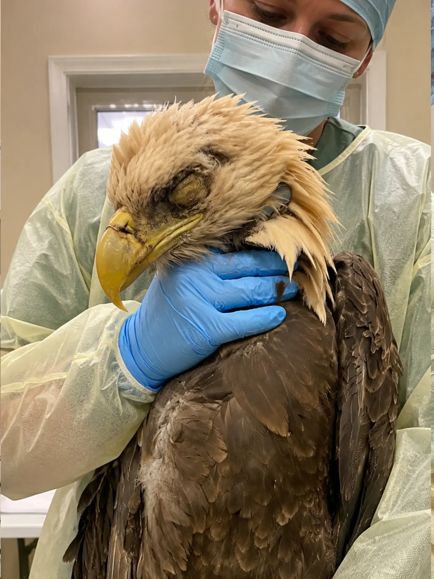
The combination has left the eagle with what its veterinary team describes as an extremely guarded prognosis.
“At this point, it is too early to predict the outcome with confidence,” said Dr. Rebecca Lawson, a wildlife veterinarian involved in the eagle’s care at a licensed rehabilitation center in western Pennsylvania. “There are several problems occurring at the same time, and we still do not know which came first.”
The eagle was discovered near Route 422 after a passerby noticed that the bird remained on or near the ground rather than flying away. It was still alive, but its posture and behavior were abnormal for a healthy adult bald eagle.
Wildlife responders were contacted, and the bird was transported for veterinary evaluation.
Once it reached the rehabilitation center, the severity of the case began to emerge.
The most obvious concern involved the head.
One eye appeared badly injured, and the eagle’s neurological responses raised concern that the trauma might extend beyond the visible tissues surrounding the eye. Diagnostic imaging later showed a fracture involving the skull.
Based on the condition of the tissues and the appearance of the injuries, clinicians estimated that the traumatic event may have occurred approximately three to five days before the bird was found.
But the more veterinarians examined the eagle, the less likely it seemed that those few days explained everything they were seeing.
The bird was unusually thin.
Its plumage was rough and poorly maintained.
Testing also revealed a substantial intestinal parasite burden.
Then bloodwork identified another major problem.
The eagle had a dangerously elevated blood lead concentration.
That finding introduced a much more complicated possibility: the bird may already have been physiologically and neurologically compromised before whatever event fractured its skull.
“The trauma itself looks several days old, but I suspect this eagle had been compromised for longer than that,” Lawson said. “The body condition is poor, the plumage is rough, and the intestinal parasite burden is substantial. When you add a high blood lead level to that picture, it becomes very difficult to separate what happened first from what happened afterward.”
Found Near a Busy Pennsylvania Highway
Butler County lies north of Pittsburgh and includes a mixture of wooded areas, farmland, streams, reservoirs, suburban development, and major road corridors.
Bald eagles are increasingly seen throughout Pennsylvania, particularly near waterways and areas where food is available.
Seeing an eagle along a road is therefore not necessarily unusual.
Finding one unable to leave is.
A healthy adult bald eagle will generally create distance when people approach. Even an eagle feeding on carrion near a roadway typically retains the ability to move or fly away when disturbed.
The bird found near Route 422 did not behave normally.
It appeared weak, remained low, and did not demonstrate the strong escape response expected from a healthy wild eagle.
That alone was enough to justify rescue.
The bird was carefully captured and transported in a secure carrier, limiting unnecessary movement in case fractures or neurological trauma were present.
By the time it reached the wildlife center, staff were preparing for what initially appeared to be a severe trauma case.
They soon discovered they were dealing with much more.
The Head Injury Was Immediately Concerning
The eagle’s facial and ocular condition drew attention during the first examination.
One eye showed evidence of significant trauma.
The tissues surrounding it were abnormal, and clinicians needed to determine whether the injury involved only the external structures or whether deeper ocular damage was present.
That distinction is especially important in a bald eagle.
Vision is central to virtually every aspect of its survival.
Bald eagles rely on precise visual information to locate food, judge distance, maneuver during flight, land accurately, identify hazards, and interact with other birds.
A serious eye injury can therefore affect release potential even if every other injury eventually heals.
But the eye was not the only concern.
The bird’s head posture and neurological presentation prompted the team to investigate possible cranial trauma.
Diagnostic imaging was performed.
The results confirmed a skull fracture.
A Skull Fracture Estimated to Be Several Days Old
Veterinarians cannot always determine exactly when a wildlife injury occurred.
There is often no witness.
The patient cannot provide a history.
Instead, clinicians work backward from the condition of wounds, swelling, tissue response, and other physical findings.
In this case, the head trauma appeared to be at least several days old.
The team estimated that the traumatic event may have occurred approximately three to five days before the eagle was discovered.
That estimate provided part of the timeline.
It did not explain the entire medical picture.
A bald eagle can deteriorate quickly after severe head trauma, but the degree of weight loss and overall poor condition suggested the possibility of a longer process.
“The injury tells us something happened several days ago,” Lawson said. “It does not necessarily tell us that the eagle was healthy before that happened.”
That difference became central to the case.
The Eye Injury Complicates the Prognosis
The injured eye remains one of the most important unknowns.
During the acute phase of trauma, swelling, inflammation, bleeding, and pain can make it difficult to determine exactly how much vision remains.
The veterinary team must first stabilize the eagle and allow some of the acute changes to subside.
Only then can clinicians begin to assess the long-term visual prognosis with greater confidence.
If the eye remains functional, that would improve the eagle’s chances of eventual release.
If the injury causes substantial permanent visual impairment, the situation becomes more complicated.
A wild bald eagle needs reliable depth perception and spatial awareness.
It must judge the distance to a perch.
It must track moving prey.
It must avoid branches, vehicles, utility infrastructure, and other obstacles.
Even a bird that can technically fly may not be capable of surviving safely if vision is severely compromised.
For now, the veterinary team is not making a final determination.
The eye is being treated and monitored while the bird’s neurological condition is assessed alongside it.
Bloodwork Revealed a Second Major Problem
The eagle’s head injuries would have been serious enough on their own.
Blood testing revealed an additional threat.
Lead.
The eagle had an elevated blood lead concentration significant enough to become a major clinical concern.
Lead toxicosis is well recognized in bald eagles and other scavenging raptors.
Depending on the level of exposure and the amount absorbed, lead can affect the nervous system, gastrointestinal function, blood production, muscle control, and overall physical condition.
Affected birds may become weak.
They may lose coordination.
They may have difficulty standing or flying.
Their reaction times can slow.
Appetite may decrease.
Severely affected eagles can remain grounded even when no major orthopedic injury is visible.
Those signs overlap with several features observed in this bird.
That overlap raised a difficult question.
Was the eagle injured first and then exposed to lead?
Or had lead poisoning weakened the bird enough to contribute to the traumatic event?
No one yet knows.
Could Lead Poisoning Have Contributed to the Trauma?
One medically plausible sequence is that the eagle was already suffering neurological effects from lead exposure before the head injury occurred.
A weakened eagle may fly poorly.
Its coordination may be impaired.
Its ability to avoid obstacles can decrease.
Reaction time may slow.
That could increase the risk of collision with a vehicle, structure, tree, or another object.
But plausibility is not proof.
Veterinarians cannot establish from the blood result alone that lead poisoning caused the skull fracture.
The fact that the bird was found near Route 422 makes a vehicle strike one possibility.
But no one witnessed such an event.
Other forms of blunt trauma could produce similar injuries.
“There is no eyewitness account, so we have to be careful,” Lawson said. “A vehicle collision is certainly something we consider because of where the eagle was found, but we cannot say that is what happened.”
For now, the mechanism of trauma remains undetermined.
The Bird’s Thin Condition Suggests a Longer Decline
One of the strongest reasons veterinarians suspect the eagle had been struggling before the head injury is its body condition.
The bird was notably thin.
Raptors can lose weight rapidly during illness, but substantial loss of muscle and body reserves generally suggests that normal feeding or physiological function has been impaired for more than a very short period.
Veterinarians evaluate body condition in birds by examining the muscle and tissue surrounding the keel and other anatomical landmarks.
Feathers can make a bald eagle appear larger than it actually is.
Underneath the plumage, a bird may have significantly reduced muscle mass.
That appears to be part of the concern in this case.
The eagle did not simply arrive with an injured head.
It arrived depleted.
That makes recovery harder.
Healing a skull injury requires energy.
Clearing lead from the body requires physiological resilience.
Rebuilding muscle requires adequate nutrition.
A patient beginning treatment with limited reserves has less margin for additional stress.
The Plumage Also Raised Questions
The eagle’s feathers were another clue.
Healthy bald eagles invest substantial time in preening and maintaining their plumage.
Feathers are not decorative.
They are essential for insulation, waterproofing, aerodynamic control, and protection.
A bird that has been chronically sick, neurologically impaired, or unable to maintain normal behavior may stop caring for its feathers effectively.
The eagle’s plumage appeared rough and poorly maintained.
That alone cannot determine how long the bird had been ill.
But when combined with its thin body condition and other findings, it reinforced the suspicion that the decline may have started before the more recent traumatic event.
The image recorded during veterinary care reflects part of that condition.
The adult eagle remains alive while a veterinary professional supports it during examination. The head feathers appear disordered, and one eye is clearly abnormal.
The image itself cannot diagnose lead poisoning or establish a skull fracture.
Those findings came from blood testing and diagnostic imaging.
But it does show a bird requiring significant professional handling and medical evaluation.
A Heavy Intestinal Parasite Burden Added Another Stressor
Testing also revealed a substantial intestinal parasite burden.
Wild animals commonly carry some parasites, and their presence does not automatically mean disease.
The significance depends on the species involved, the number present, and the condition of the host.
A healthy wild bird may tolerate a modest parasite burden without obvious problems.
A severely compromised bird may not.
In this eagle, veterinarians considered the parasite burden an additional physiological stressor rather than the primary explanation for the entire case.
The bird was already thin.
It was dealing with head trauma.
It had elevated lead in its bloodstream.
Adding a significant parasite load further complicated recovery.
Parasites can reduce nutritional efficiency and place additional demands on a body already trying to heal.
The medical team therefore has to address several problems simultaneously without overwhelming an already weakened patient.
The Source of the Lead Is Unknown
Identifying lead poisoning does not automatically identify the source.
Bald eagles are opportunistic feeders.
They consume fish, birds, mammals, and carrion.
One recognized pathway of lead exposure occurs when scavenging birds ingest fragments of lead embedded in animal remains.
Small particles can be consumed unknowingly with tissue.
Other pathways may also exist.
But in this case, veterinarians cannot say where the exposure occurred.
The eagle may have traveled substantial distances before becoming too weak to continue.
Where it was found does not necessarily correspond to where it encountered the lead.
That is why the reporting must remain precise.
The eagle has elevated lead levels.
The source has not been determined.
Treatment Has to Address Two Emergencies at Once
The rehabilitation team is now managing both traumatic injury and toxic exposure.
For the head trauma, priorities include pain control, stabilization, neurological monitoring, eye care, and restricted activity.
For the lead exposure, treatment may include chelation therapy, supportive fluids, nutritional care, and repeat blood testing.
Chelating medications work by binding to lead and helping the body eliminate it.
The process takes time.
A single treatment does not instantly restore neurological function.
The veterinary team must watch both the blood values and the bird itself.
Does lead concentration decrease?
Does posture improve?
Does the bird become more alert?
Can it stand more consistently?
Does appetite return?
Is coordination improving?
The laboratory data and clinical behavior have to move in the same direction.
Nutritional Support Is Especially Important
Because the eagle entered care in poor body condition, nutrition is one of the most important parts of treatment.
A severely weakened bird may not immediately eat on its own.
Pain, neurological impairment, stress, eye trauma, and general illness can all suppress feeding behavior.
The team must determine whether the eagle can swallow normally and whether supportive nutritional intervention is required.
Weight will be followed closely.
Even small additional losses matter in an already thin patient.
The goal is eventually to see the bird maintain or gain weight through normal independent feeding.
That milestone would signal improvement in more than one system.
It would suggest better appetite, stronger neurological function, and enough overall stability to support recovery.
The First 24 to 72 Hours Are Critical
With a case this complicated, clinicians watch early changes very closely.
A bird can improve.
It can also deteriorate rapidly.
Neurological status is particularly important after head trauma.
Staff monitor the eagle’s level of awareness, head position, balance, eye responses, and ability to stand.
Any seizure-like activity or worsening coordination would be concerning.
The injured eye is also reassessed for changes in swelling and tissue viability.
At the same time, the medical team monitors hydration, appetite, body weight, and response to lead treatment.
If the eagle becomes more responsive and begins behaving more normally, those would be encouraging signs.
But improvement over one day would still not provide enough information to predict eventual release.
“Very Poor” Does Not Mean the Outcome Is Already Decided
The veterinary team has described the eagle’s overall condition as very poor.
That reflects the seriousness of the findings.
It does not mean the bird has no chance.
Wildlife patients can sometimes recover from combinations of injuries that initially appear severe.
The difficulty is that several independent problems have to improve.
The skull fracture must heal without severe neurological consequences.
The eye injury must stabilize.
The lead burden must fall.
The bird must regain appetite and body condition.
The parasite burden must be managed appropriately.
Only after those immediate medical issues are under control can the team begin asking whether the eagle remains capable of surviving independently.
That is why the prognosis is best described as extremely guarded.
There are simply too many unanswered questions to make a confident prediction.