A bald eagle rescued from a canal in Cape Coral, Florida, appeared surprisingly alert and physically stable when it arrived for veterinary care, but a later flight assessment and diagnostic imaging revealed an injury that could not be seen from the outside: a fracture of the coracoid, one of the major bones of the avian shoulder girdle and a structure essential to normal flight.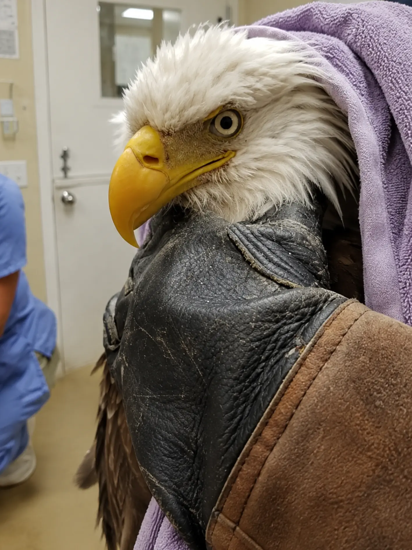
The adult eagle had been pulled from the water only a day earlier after rescuers saw it struggling in one of Cape Coral’s canals. Once safely on shore, the bird remained upright and responsive, raising hope that it might simply be exhausted or temporarily compromised after spending time in the water.
Rescuers initially gave the eagle an opportunity to recover.
But it did not fly away.
That single detail changed the direction of the case.
A healthy adult bald eagle would ordinarily be expected to put distance between itself and approaching people once it had recovered enough to stand. When this bird remained grounded despite being conscious and aware of its surroundings, rescuers began to suspect that something more significant was preventing it from leaving.
The eagle was secured for transport and taken to a staging point before being transferred to a licensed wildlife hospital in Southwest Florida.
At the clinic, however, the mystery deepened.
The eagle stood upright.
Its eyes were open and responsive.
There was no obvious open wound, no visibly displaced wing, and no dramatic external injury that immediately explained why such a powerful bird had been unable to leave the canal.
Only after the veterinary team evaluated the eagle’s movement in a controlled outdoor enclosure did the hidden problem become clearer.
The bird was reluctant to use its left wing normally.
Radiographs were then taken.
The images revealed a coracoid fracture.
For the rehabilitation team, the finding finally provided an explanation for a case that had looked deceptively mild on the surface.
The eagle could stand.
It could watch the room.
It could appear remarkably composed.
But one of the structures that allowed its wings to withstand the forces of flight was broken.
A Rescue From One of Cape Coral’s Canals
Cape Coral is defined in part by water.
The Southwest Florida city contains an extensive network of canals that run through residential neighborhoods and connect with larger waterways throughout the region. Those canals attract fish, wading birds, waterfowl, and other wildlife, making encounters with native animals relatively common.
Bald eagles also occur throughout Florida.
The state supports a substantial breeding population, and adults may be seen around lakes, rivers, reservoirs, wetlands, coastal habitats, and developed areas where suitable food and nesting habitat remain available.
Seeing a bald eagle near water is therefore not unusual.
Seeing one unable to get out of the water is.
Responders became concerned when the eagle was observed struggling in a canal rather than simply feeding or bathing.
Bald eagles can enter water while attempting to capture fish, and they are capable of moving through the water using their wings under some circumstances. But prolonged difficulty at the surface can indicate exhaustion, injury, or another condition preventing normal flight.
The bird needed assistance.
Rescuers were able to reach it and bring it safely onto shore.
At that moment, the most immediate danger had passed.
The eagle was no longer struggling in the canal.
What remained unclear was why it had ended up there.
Rescuers Waited for the Bird to Leave
Once on land, the eagle was given time to recover from the stress of the rescue.
Wet plumage can make a bird temporarily heavier and reduce insulation. An animal that has struggled in water may also be exhausted even if it has no major injury.
If those were the only problems, rest might have been enough.
The eagle remained alert.
That was encouraging.
But it did not take off.
Wildlife responders pay close attention to that type of behavior because a bald eagle’s response to human proximity is itself an important indicator of function.
A healthy adult bird generally does not remain quietly on the ground while people are nearby.
It may walk away.
It may become defensive.
Most importantly, if it has enough room and normal flight ability, it usually leaves.
This eagle did not.
The decision was therefore made not to continue testing it at the scene.
Repeated attempts to make an injured bird fly can worsen an orthopedic problem that has not yet been diagnosed.
Instead, the eagle was contained for transport.
From Rescue Site to Veterinary Care
Transporting an adult bald eagle requires careful handling.
Even a bird that is weak or injured can retain significant strength in its feet and talons.
Those feet are designed to capture and hold prey and can cause severe injuries to anyone who attempts to handle the bird without appropriate training.
At the same time, rescuers have to protect the eagle from itself.
An injured raptor may struggle instinctively during capture.
If a wing or shoulder is damaged, uncontrolled movement can turn a stable injury into a more severe one.
The bird was therefore placed in a secure transport container and moved to a staging point while arrangements were made for veterinary evaluation.
Keeping the eagle contained also reduced the temptation to repeatedly “test” whether it could fly.
That restraint would prove important.
The injury later identified on radiographs was exactly the kind that could have been aggravated by forcing the bird into strenuous wingbeats too soon.
The Eagle Looked Better Than Expected at the Clinic
When the eagle arrived at the wildlife hospital, veterinary staff saw a bird that did not immediately resemble a patient with a significant fracture.
It was upright.
Its eyes were open.
It tracked movement around the room.
Its posture was reasonably controlled.
No major external wound was obvious.
The wings did not display the dramatic deformity commonly associated with certain fractures of the outer wing.
That appearance could easily have created false reassurance.
Instead, the team considered the history.
The eagle had been found struggling in water.
It had been rescued.
And after being given an opportunity to recover, it still had not flown away.
Those facts could not be ignored simply because the bird now looked stronger.
Wildlife medicine often depends on combining what is seen in the examination room with what happened before the patient arrived.
In this case, the behavior at the canal was one of the most important pieces of medical information.
A Complete Physical Examination
Veterinary staff began by assessing the eagle systematically.
Respiration was evaluated.
The eyes and pupils were examined.
The beak and head were checked for evidence of trauma.
Both wings were inspected and compared.
The shoulders, elbows, wrists, and major wing bones were assessed for swelling, asymmetry, abnormal movement, or pain.
The legs and feet were also examined.
A bird that has fallen into water could have suffered trauma elsewhere in the body before entering the canal.
Neurological function mattered as well.
The team needed to know whether the inability to fly might be related to impaired coordination rather than a purely orthopedic problem.
Hydration and body condition provided additional information about how long the bird may have been compromised.
Nothing on the initial surface examination produced an obvious answer.
That did not mean the bird was normal.
It meant the problem might be located somewhere more difficult to evaluate externally.
Why Shoulder-Girdle Injuries Can Be Hidden
Bird anatomy makes certain injuries particularly difficult to recognize without imaging.
The shoulder is supported by a complex arrangement of bones and soft tissues that differ substantially from those of mammals.
Among the key bones are the scapula, clavicles, and coracoids.
The coracoid extends between the shoulder region and the sternum and forms an important component of the structural system that stabilizes the shoulder during flight.
Unlike fractures farther down the wing, a coracoid fracture may not create a clearly dangling or visibly deformed wing.
The bone lies deep within the body, surrounded by muscle and other structures.
An eagle with this injury may still sit upright.
It may still move its head normally.
It may even reposition itself within an enclosure.
What becomes difficult is generating the forces necessary for normal flight.
That difference explains why functional assessment became so important in this case.
The Outdoor Flight Assessment Revealed the Problem
Once the eagle had been stabilized and the veterinary team considered a controlled assessment appropriate, the bird was evaluated in an outdoor enclosure.
The purpose was not to force sustained exercise.
It was to observe whether the eagle used both wings normally under conditions where it had enough room to attempt natural movement.
That was when the asymmetry became more apparent.
The eagle was reluctant to use the left wing normally.
Its wing action did not match what clinicians would expect from a healthy adult bald eagle.
The problem was subtle enough that it had not been obvious while the bird was standing quietly in the treatment area.
Under the mechanical demand of attempted flight, however, the abnormality became much harder to miss.
The team stopped there.
The next step was imaging.
X-Rays Provided the Missing Answer
Radiographs revealed a fracture of the coracoid.
That finding transformed the understanding of the case.
The eagle had not simply been exhausted after struggling in the canal.
A major structural component of its shoulder girdle had been injured.
The fracture also explained why the bird could look comparatively healthy while resting but fail when asked to fly.
Standing places very different mechanical demands on the body than flight.
During a wingbeat, the shoulder experiences powerful forces.
The coracoid helps brace that system.
When it is fractured, the bird may be unable to generate or withstand normal flight forces even if the rest of the wing looks externally intact.
The X-ray therefore provided an anatomical explanation for the clinical signs responders had seen from the beginning.
What the Coracoid Does
In birds, the coracoid is one of the key structural elements connecting the shoulder to the sternum.
It contributes to the rigid framework required to support flight.
During the downstroke, large flight muscles generate substantial force.
The shoulder girdle must resist deformation while allowing controlled movement of the wing.
The coracoid plays an important role in maintaining that stability.
In a large bird such as a bald eagle, those forces are considerable.
A fracture can reduce the bird’s ability to use the wing effectively, creating pain, weakness, or altered mechanics.
Depending on the location and severity of the break, the bird may avoid fully extending or loading the affected wing.
That can lead to asymmetrical flight or complete reluctance to take off.
For an animal whose survival depends on strong and precise flight, the implications are serious.
The Canal May Have Been the Result, Not the Cause
One of the most important questions was whether the eagle had injured itself in the canal or had entered the water because it was already injured.
The fracture makes the second possibility plausible.
If the coracoid had been damaged before the bird reached the water, the eagle may have lost flight efficiency or been unable to regain altitude after descending.
It could then have ended up in the canal and found itself unable to escape.
But the fracture alone cannot reconstruct that sequence with certainty.
Veterinarians cannot determine from an X-ray exactly where the original injury occurred.
A collision with an object might cause such trauma.
A hard impact during another event could also be responsible.
Without a witness, the precise mechanism remains unknown.
The safest conclusion is that the eagle sustained significant blunt trauma, and the resulting coracoid fracture likely contributed to its inability to fly normally.
Anything more specific would be speculation.
Why the Lack of an External Wound Mattered
The case demonstrates why external appearance can be misleading in wildlife trauma.
Many people expect a serious injury to look dramatic.
Blood.
A visibly broken wing.
An obvious wound.
This eagle had none of those features prominently visible when it arrived.
Instead, it was alert enough that an observer might reasonably have wondered why it needed to remain in care at all.
That is exactly where history and functional testing become essential.
The inability to leave the canal was abnormal.
The reluctance to use the left wing was abnormal.
Those signs justified further investigation.
Without radiographs, the injury might have remained hidden until the bird attempted more demanding flight.
In the wild, that failure could have been fatal.
Treatment Begins With Restriction, Not Exercise
Once the coracoid fracture was identified, the rehabilitation plan had to protect the shoulder from excessive load.
That meant restricting activity.
For an eagle, this can seem counterintuitive.
The ultimate goal is flight, yet the first step toward that goal may be preventing flight entirely.
The reason is mechanical.
Every strong wingbeat transmits force through the shoulder girdle.
If the fracture is not stable, repeated attempts to fly could interfere with healing or worsen displacement.
The bird therefore needs a period in which the damaged bone can begin to stabilize without being subjected to full flight forces.
Housing during this stage is intentionally controlled.
The eagle needs enough space to stand comfortably and reposition itself, but not so much that it repeatedly launches into strenuous flight.
Pain Management and Supportive Care
Pain control is also important.
Wild birds often conceal signs of discomfort, but that does not mean painful injuries are not affecting them.
A coracoid fracture may cause pain during movement even when the bird appears relatively calm at rest.
Appropriate analgesia can improve welfare and reduce unnecessary stress.
Veterinary staff also continue monitoring appetite, hydration, posture, wing position, and overall responsiveness.
A bird recovering from a fracture still has ordinary physiological needs.
If it stops eating or becomes increasingly inactive, those changes may signal that additional problems are developing.
The rehabilitation team must therefore manage the entire patient rather than focusing only on the broken bone.
Follow-Up Imaging Will Matter
Coracoid healing takes time.
Repeat radiographs may be used to evaluate whether the fracture is stabilizing and whether new bone formation is progressing appropriately.
Alignment is important.
So is the bird’s clinical response.
An X-ray that looks better is encouraging, but the medical team also wants to see improved comfort and increasingly normal movement.
In some cases, a fracture may heal structurally yet leave the shoulder with reduced function.
That is particularly important in flying animals.
A person might tolerate a small loss of shoulder mobility during ordinary daily life.
A bald eagle has much less room for compromise.
Bone Healing Is Not the Same as Flight Recovery
This distinction will become central later in the eagle’s rehabilitation.
A fracture may show evidence of healing while the bird is still not ready to fly.
Restricted movement causes muscle deconditioning.
The injured side may lose strength.
Scar tissue can affect range of motion.
The bird may also develop compensatory habits, relying more heavily on the healthy wing during movement.
Those issues have to be addressed gradually.
Once imaging and examination show that the fracture can safely tolerate greater activity, the rehabilitation plan will shift.
The eagle will move from protection toward controlled rebuilding.
Reconditioning the Shoulder
The next stage will focus on restoring function without overwhelming healing tissues.
Initially, staff may observe ordinary wing extension and movement.
They will compare both sides.
Does the eagle stretch the left wing normally?
Does it hold both wings symmetrically?
Is there still hesitation?
As strength returns, the bird can be given progressively more space.
The goal is to restore the entire flight system, not simply the bone.
Muscles need conditioning.
Joints need mobility.
The bird must regain confidence using the previously painful side.
These changes occur over time.
Rushing them creates a risk of reinjury.
The Flight Enclosure Will Eventually Provide the Real Test
If the fracture heals appropriately, the eagle may eventually move into a large flight enclosure.
This stage is crucial because it recreates the physical demands that cannot be evaluated in a small treatment room.
Staff will watch the first takeoff carefully.
A healthy bald eagle should generate strong, symmetrical wingbeats.
The bird should gain elevation efficiently.
If the left wing remains weaker, the eagle may tilt or fail to climb normally.
Sustained flight provides additional information.
Does the bird maintain altitude?
Does it tire quickly?
Are the wingbeats equal throughout the flight rather than only at takeoff?
Then come turns.
An eagle needs to maneuver accurately in both directions.
Landing is equally important.
The bird must slow, extend the feet, judge the perch, and absorb the final impact while maintaining balance.
Why One Successful Flight Will Not Be Enough
A recovering eagle may surprise its caregivers with one good flight.
That does not automatically make it releasable.
Wild life demands repetition.
An eagle may make many flights in a single day.
It may travel considerable distances between feeding and roosting areas.
It may encounter wind.
It may need to climb rapidly or change direction to avoid other birds.
A shoulder that tolerates one short flight but becomes painful or weak after repeated effort is not ready for release.
Rehabilitators will therefore evaluate endurance as well as capability.
The eagle needs to demonstrate that the repaired shoulder can withstand sustained use.
The Rest of the Bird Must Still Be Normal
Even though the coracoid fracture provides a strong explanation for the inability to fly, the veterinary team still has to ensure no other problem limits release.
The eyes must function normally.
The feet and talons need to provide secure grip.
Neurological coordination must remain intact.
The bird must maintain healthy body condition.
It must feed independently.
And it must remain appropriately wary of people.
Wildlife rehabilitation does not release a collection of healed injuries.
It releases a whole animal.
Everything essential to independent survival must work together.
Why the Image Shows a Bird That Looks So Healthy
The photograph taken during veterinary care demonstrates exactly why this case was initially deceptive.
The eagle is alert.
Its head is upright.
The eye is open and attentive.
The beak looks externally normal.
The bird is being carefully restrained in a towel while experienced personnel handle it.
Nothing in that image alone reveals a fractured coracoid.
That is the point.
The injury lies deep inside the shoulder girdle.
It is not something an observer can diagnose by looking at the bird’s feathers.
Only the combination of history, functional assessment, physical examination, and radiographs revealed the true problem.
Rescuers Were Right Not to Force Another Flight
The case also validates the decision made immediately after the canal rescue.
When the eagle did not fly away, responders did not repeatedly chase it or throw it into the air in an attempt to see whether it “really” could fly.
That could have placed unnecessary stress on a fractured shoulder.
Instead, the abnormal behavior was treated as meaningful.
The bird was transported for evaluation.
That cautious approach gave the medical team the opportunity to identify the fracture before the eagle was subjected to repeated strenuous flight attempts.
In wildlife rescue, restraint can sometimes be as important as intervention.
The Prognosis Remains Guarded but Hopeful
A coracoid fracture is serious because of the bone’s importance to flight.
It is not automatically incompatible with recovery.
The outlook depends on several factors, including the exact fracture configuration, alignment, stability, associated soft-tissue injury, and how the bird responds during healing.
If the bone remains well positioned and the surrounding structures recover appropriately, the eagle may regain full function.
If the fracture heals poorly or leaves significant restriction in shoulder mechanics, release may become more difficult.
That uncertainty is why the prognosis remains cautious.
The team has a clear diagnosis.
The outcome will depend on function.
Why Release Standards Must Be Strict
A bald eagle returned to the wild will receive no further accommodations.
There will be no controlled enclosure.
No padded surface.
No easy perch positioned nearby.
No staff member watching whether the left wing begins to tire.
The bird will need to manage every aspect of survival independently from the moment it leaves rehabilitation.
That means flight must be reliable.
Not adequate.
Not almost normal.
Reliable.
The eagle must be capable of taking off from uneven terrain, climbing, maneuvering, landing, and repeating those movements without pain or significant weakness.
A bird with only partial shoulder recovery may survive temporarily after release but remain unable to compete effectively for food or escape danger.
That is why rehabilitation teams are careful.
The objective is not simply to open a carrier.
It is to ensure the bird has a realistic chance after the carrier is opened.
A Canal Rescue Became an Orthopedic Case
The story began with an eagle in the water.
That naturally made the canal itself appear to be the central problem.
Once the bird was pulled out, however, the true challenge emerged.
The canal was where the eagle was found.
The coracoid fracture was why it could not simply leave.
That distinction changes the entire medical narrative.
Water rescue was the first intervention.
Orthopedic rehabilitation became the longer one.
The Final Answer Will Come From the Eagle
Veterinarians can take radiographs.
They can monitor bone healing.
They can control pain.
Rehabilitators can create increasingly demanding opportunities for movement.
But eventually, the eagle itself has to provide the final evidence.
The shoulder must work.
The wings must move together.
The bird must fly.
If the eagle eventually launches strongly from a rehabilitation perch, gains altitude, turns accurately, lands with control, and repeats those behaviors without weakness, the medical team will have evidence that the hidden fracture has truly been overcome.
Until then, recovery remains incomplete.
From the Canal Toward a Possible Return to the Wild
A day before reaching the wildlife hospital, the bald eagle had been struggling in a Cape Coral canal, unable to escape under its own power.
Rescuers pulled it from the water and waited.
The bird remained alert.
It stood.
It looked better.
But it did not fly.
That refusal to leave became the first important clue.
At the hospital, the mystery continued because the eagle still looked unexpectedly healthy.
Only when the bird was evaluated under the demands of flight did the problem become visible.
The left wing was not being used normally.
Radiographs then revealed the fracture hidden deep within the shoulder girdle.
The coracoid injury finally connected the pieces.
Why the bird struggled in the canal.
Why it stayed on the ground afterward.
Why it looked stronger than its actual flight capacity suggested.
And why allowing it to leave without a complete evaluation could have been dangerous.
For now, the eagle remains under professional care.
The fracture needs time.
The shoulder needs protection.
Later, the wing will need strength.
And eventually, if healing progresses well enough, the bird will face the same test that first exposed the problem: flight.
Standing upright in an examination room, the eagle gave few outward clues that anything was broken.
It was only when the bird tried to fly that the hidden injury revealed itself.
The X-rays finally showed why an adult bald eagle that appeared so alert and composed had been unable to escape a Florida canal.
Whether that same bird can one day return to the open skies of Southwest Florida will depend not merely on whether the fracture heals on an X-ray, but on whether the shoulder can once again withstand the extraordinary forces of wild flight.