Tonight, an injured adult bald eagle is receiving emergency care at a licensed raptor rehabilitation center in northern Minnesota after arriving shortly before dark with signs of physical trauma, dehydration and reduced appetite.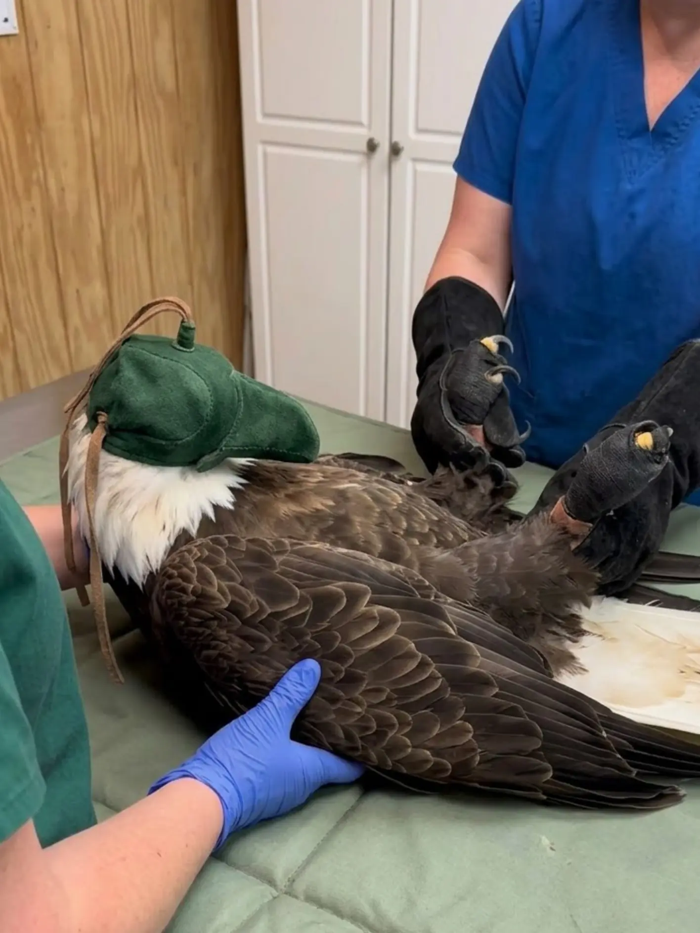
The bird was conscious and responsive on admission, but it was clearly uncomfortable and was not moving with the confidence expected from a healthy adult bald eagle. Staff immediately began a full physical examination, checking the wings, shoulders, legs, feet, chest, spine and neurological responses before deciding how aggressively the bird could safely be handled.
The first several hours of treatment were deliberately methodical.
The eagle was examined.
Fluids were administered to support hydration and circulation.
Small pieces of food were offered because the bird was not yet eating normally on its own.
And once the conventional veterinary assessment had ruled out any obvious unstable injury that would make additional manipulation unsafe, the eagle also received a gentle musculoskeletal evaluation from a veterinarian trained in animal manual therapy.
One part of that evening’s care may look uncomfortable on video: staff using forceps to hold small pieces of meat near the bird’s beak.
The rehabilitation team emphasized that the instrument is not being pushed into the eagle or used to restrain tissue.
“The forceps can look harsher on camera than they actually are,” one rehabilitator explained. “I am only using them to hold the food. The tips are not piercing the mouth, the throat or any part of the bird. It is a controlled technique used to place a small piece of meat where the eagle can safely take and swallow it.”
For wildlife staff, that distinction matters.
Assisted feeding is not simply about getting calories into an animal.
It is also a clinical test.
Can the eagle focus on the food?
Can it open its beak normally?
Can it grasp or accept the piece?
Can it swallow safely?
Those small responses can tell veterinarians a great deal about the bird’s neurological status, comfort level and ability to begin recovering from the stress of rescue.
For now, the eagle remains under close observation.
The prognosis is guarded while the team waits to see how it responds overnight and whether its posture, appetite and overall mobility improve.
The First Examination Began With Safety
Handling any adult bald eagle requires planning.
Even a seriously injured bird may retain tremendous strength in its feet.
The talons remain one of the greatest immediate risks to anyone conducting an examination.
For that reason, the rehabilitation team used coordinated restraint.
One staff member controlled the legs and feet with protective equipment.
Another supported the bird’s body and wings.
The head was covered with a soft hood to reduce visual stimulation.
That hood does not anesthetize the eagle and is not intended to interfere with breathing.
It simply reduces the amount of movement and environmental activity the bird can see during the examination.
For a wild eagle, being restrained in a brightly lit room while several people work around it can be extremely stressful.
Reducing visual input can help lower unnecessary struggling.
The goal is always to use the least restraint necessary to complete the examination safely.
The image taken during the evening evaluation reflects this approach.
The eagle is lying on a padded examination surface.
Its head is covered.
The feet are being controlled by staff wearing protective gauntlets.
Another team member supports the body and wing.
The scene may look highly controlled because it has to be.
A single uncontrolled kick from an adult bald eagle can cause a serious puncture injury.
A Full Physical Examination Came Before Everything Else
Before deciding what treatments were needed, the veterinary team worked through the bird systematically.
The eyes were checked.
The beak and oral cavity were examined.
The neck was palpated.
The shoulders were compared.
Each wing was evaluated from the shoulder through the humerus, elbow, radius and ulna, wrist and distal flight feathers.
The keel and pectoral muscles were assessed for bruising, asymmetry or loss of condition.
The pelvis, legs and feet were examined for swelling, abnormal range of motion and pain.
Neurological responses were also monitored.
Could the eagle move both feet?
Were the toes functioning normally?
Did the bird react appropriately to touch?
Was head control normal?
Were the pupils symmetrical?
Did the bird appear mentally alert?
This type of examination is particularly important when the circumstances surrounding an injury are not fully known.
A grounded eagle may have been struck by a vehicle.
It may have collided with a structure.
It may have suffered trauma during a territorial encounter.
It may have been weakened by illness before the injury occurred.
Without a witness, the medical team has to work from the physical evidence.
At this stage, staff were not assigning a specific cause.
They were trying to determine what had been damaged and what the bird needed immediately.
Dehydration Was One of the Early Concerns
Wildlife patients often arrive after spending an unknown amount of time without access to water or normal food.
Stress also increases physiological demand.
An eagle that has been injured, unable to move normally or exposed to poor weather may become dehydrated relatively quickly.
The veterinary team assessed hydration based on the physical examination and overall condition.
Fluids were then administered to support hydration and circulation.
The exact route depends on the patient.
In some cases, fluids may be given intravenously.
In others, subcutaneous administration may be appropriate.
The purpose is supportive.
Fluid therapy does not heal a muscle strain.
It does not repair a broken bone.
It does not treat every cause of weakness.
What it does is improve the physiological environment in which the rest of the body has to recover.
For a bird under stress, that matters.
Why Fluids Are So Important Early in Wildlife Care
Hydration affects circulation.
It affects kidney function.
It affects how efficiently nutrients and medications are distributed.
It affects recovery from prolonged exertion.
A dehydrated animal can look more weak, more lethargic and less responsive than the underlying injury alone would predict.
That is why stabilization often begins with the simplest interventions.
Warmth.
Quiet.
Fluids.
Pain control.
Nutrition.
Only after those basic needs are addressed can clinicians accurately interpret what remains abnormal.
The eagle received supportive fluid therapy while staff continued the rest of the examination.
The Bird Was Not Yet Eating Normally
Another immediate concern was appetite.
Wild raptors frequently refuse food during the early stages of hospitalization.
Stress is one reason.
Pain is another.
An unfamiliar environment can suppress feeding.
A bird that has recently been restrained may simply be too alert to human presence to eat voluntarily.
But prolonged refusal to eat creates problems of its own.
Adult bald eagles are large birds with substantial metabolic needs.
A patient already expending energy on healing cannot afford unnecessary nutritional decline.
The rehabilitation team therefore began offering small, appropriate pieces of meat.
The goal was not to force a full meal into the bird.
It was to assess whether it could take food safely and begin supporting nutritional intake.
The Feeding Forceps Look More Intimidating Than the Technique Actually Is
This is the part of care most likely to be misunderstood when seen in a short video.
A metal forceps or feeding instrument may look sharp simply because of its shape.
But experienced wildlife staff are not using it to puncture or probe the eagle.
The instrument holds the food.
That is all.
The meat is secured at the tip.
The food is brought toward the beak in a controlled way.
The bird is given an opportunity to take or accept it.
The instrument is kept away from sensitive tissue as much as possible.
If the eagle is not swallowing normally, feeding stops.
If the bird shows signs that the technique is unsafe, the approach is reassessed.
“This is something we do carefully and very deliberately,” the rehabilitator explained. “I know it can look rough from the outside, but the forceps never need to stab or push into the bird. They are simply holding the piece of meat. The bird’s reaction and swallowing are monitored the entire time.”
Why Staff Do Not Simply Push Food Into the Mouth
Assisted feeding is not a matter of placing food as deep as possible.
That would be dangerous.
A bird with neurological impairment, severe weakness or altered swallowing may aspirate material into the respiratory tract.
That can produce serious complications.
Food therefore has to be offered in a way that allows the bird to demonstrate an effective swallow.
Small pieces are safer than large ones.
The pace is slow.
The staff member watches the throat and beak.
If the bird does not coordinate normally, feeding is stopped.
The procedure provides information while also providing nutrition.
Eating Is a Neurological Sign as Well as a Nutritional One
A bird that visually tracks food, opens the beak and swallows appropriately is showing several systems working together.
Vision.
Attention.
Jaw movement.
Tongue and throat coordination.
Swallowing.
That does not prove the bird is neurologically normal.
But it is useful information.
For this eagle, accepting at least some assisted feeding was considered more encouraging than complete refusal.
The team hoped that after several hours of rest, the bird would begin eating voluntarily.
Voluntary feeding would reduce the need for handling and offer a much stronger sign that the patient was becoming more stable.
Pain Can Suppress Appetite
One reason the eagle may have been reluctant to eat was discomfort.
Wild animals do not always show pain in obvious ways.
A bird may remain quiet.
It may hold one wing differently.
It may resist certain movements.
It may simply stop eating.
Those changes can be subtle.
Pain control is therefore part of responsible wildlife rehabilitation when trauma is suspected.
Analgesia may help the bird rest more effectively.
It can reduce stress.
It can allow more normal posture and movement.
It can also make later examinations more meaningful because staff can better distinguish true mechanical limitation from protective movement caused by pain.
Imaging May Be Needed Depending on Examination Findings
If the examination reveals focal swelling, restricted wing movement, instability or pain over a specific bone, radiographs are typically considered.
The team does not rely on external appearance alone.
Bald eagle feathers hide injuries remarkably well.
A fracture may be difficult to see beneath dense plumage.
A bird can sometimes hold an injured wing close enough to the body that the problem is not obvious until palpation.
Likewise, bruising can be extensive without a bone fracture.
Imaging is used to separate those possibilities.
In this case, the initial treatment plan remained focused on stabilization while the team evaluated whether additional diagnostics would be required.
The Musculoskeletal Evaluation Came After Conventional Veterinary Assessment
Later in the evening, the eagle also received a gentle musculoskeletal evaluation from a veterinarian trained in animal manual therapy.
This part of the care deserves careful explanation.
Manual therapy is not being used as a replacement for diagnostic imaging, pain medication, fracture management or conventional veterinary examination.
It is an adjunct.
The first priority is always to rule out injuries that would make manipulation unsafe.
An unstable fracture should not be manually adjusted.
A dislocated joint requires appropriate veterinary management.
Severe spinal trauma requires careful diagnostic assessment.
Only when those immediate concerns have been considered can a gentle manual examination be used to assess things like muscular tension, range of motion and asymmetry.
What a Gentle Manual Assessment Can Evaluate
The veterinarian may compare movement through the shoulders.
The neck can be assessed for stiffness.
Pelvic symmetry can be observed.
Muscular tension around the back and shoulder girdle can be evaluated.
Range of motion can be compared from side to side.
The goal is not to “crack” bones back into place.
The goal is to gather additional information about how the musculoskeletal system is functioning after trauma.
If a particular movement produces significant pain or resistance, the veterinarian stops.
That response may indicate that additional imaging or rest is necessary.
Manual Therapy Is Not a Substitute for Diagnosis
The rehabilitation team was careful to make this distinction.
A manual treatment should never be presented as proof that a fracture, internal injury or neurological problem has been cured.
It is one component of a larger medical plan.
The eagle’s evening care still depended on standard veterinary principles:
physical examination,
hydration support,
nutrition,
pain management,
appropriate diagnostic testing,
and observation.
The manual assessment was added only after those priorities were considered.