For a few quiet seconds after the carrier door was opened, the Bald Eagle did not move.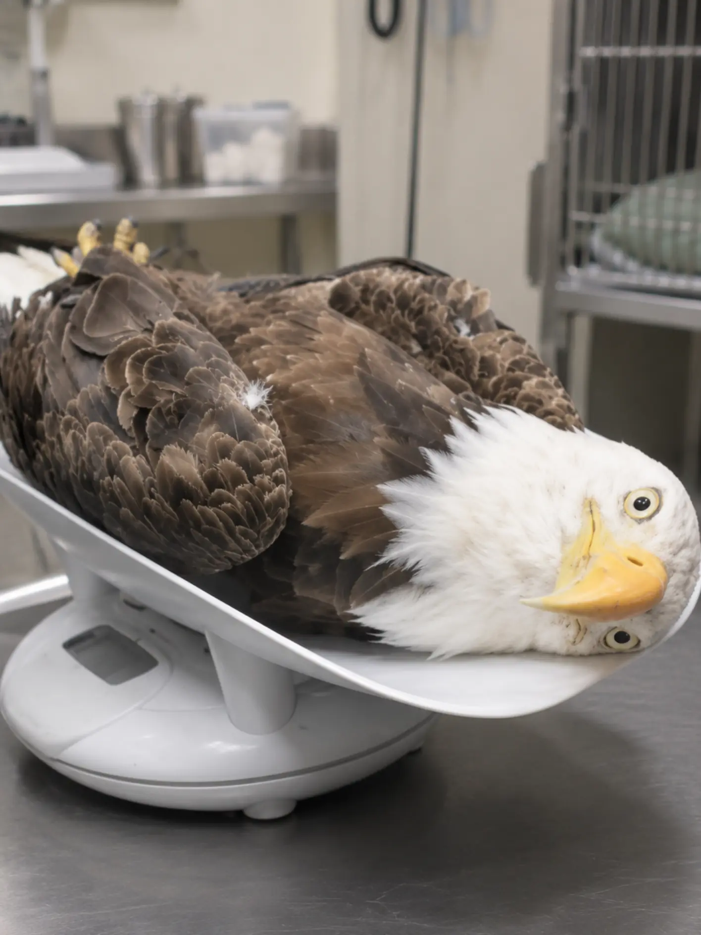
It stood at the edge of the crate, head lifted, pale eyes fixed on the open field ahead. The late-morning wind moved lightly through the grass, and the people gathered a respectful distance away fell silent. No one called out. No one stepped forward. After everything that had happened, this final decision belonged to the bird alone.
Then, almost without warning, it leaned forward.
Its wings opened in one clean, even motion — broad, steady, controlled — and the eagle launched into the air. It cleared the grass, caught the wind, and climbed. Within seconds it had crossed the field, passed over the line of trees, and disappeared toward the river corridor where it belonged.
For the rehabilitation staff watching from below, the moment carried a weight that was difficult to put into words. Just weeks earlier, that same eagle had arrived in veterinary care unable to stand normally, unable to bear pain through an injured wing, and uncertain to ever fly with full strength again. What had looked, at first glance, like a grounded raptor in distress had turned into a complex case involving fracture repair, prolonged rehabilitation, and a sobering reminder of the risks Bald Eagles still face — even decades after one of the greatest wildlife recoveries in American history.
This adult Bald Eagle had not been weakened by disease alone, nor brought down by age. It had been struck by a hunter’s shot.
And yet, through rapid reporting, expert veterinary intervention, and weeks of disciplined rehabilitation, the bird was given a second chance.
The story began near the Susquehanna River in Pennsylvania, where a local resident noticed something that did not look right. Bald Eagles are powerful, unmistakable birds. Even when perched, they project balance and control. This eagle had neither. According to the initial field report, the bird was on the ground near the edge of an open area adjacent to woodland and showed repeated but unsuccessful attempts to get airborne. Each effort ended the same way: a brief burst of motion, an uneven lift, and a collapse back to the ground.
A wildlife officer responded and found an adult Bald Eagle alert but clearly compromised. One wing appeared lower than the other. The bird could stand only with difficulty and reacted defensively when approached, as a healthy wild eagle should, but its movements were limited. There was no immediately obvious catastrophic wound from a distance, and as is often true in raptor cases, the most significant injury was not fully apparent until the bird could be safely examined under controlled conditions.
The eagle was captured and transported to the fictional Allegheny Raptor Rehabilitation Center, where intake began that same day.
One of the photographs taken shortly after admission showed the eagle lying on its side on a veterinary scale, eye open, body tense, feathers slightly disordered from transport and stress. To a casual observer, the image might have looked unusual. To rehabilitation staff, it documented an important truth about the bird’s condition at the time of intake: this was not an eagle able to hold a normal stance comfortably. Pain, instability, exhaustion, and stress had already taken a serious toll.
At the center, the case was relayed to Dr. Hannah Brooks, a veterinarian with experience in avian and wildlife trauma. By the time the call came in, much of the regular clinical day had already ended. But once she heard the details — adult Bald Eagle, possible wing fracture, potentially salvageable function if treated quickly — she returned to the facility to assist with the evaluation.
Later, Brooks would say that timing mattered.
“If a wing can still be saved for flight,” she explained, “then every hour matters. A Bald Eagle can survive the first night after injury. The bigger question is whether it will still have a realistic path back to the wild if stabilization is delayed.”
That distinction shaped the next several hours.
On physical examination, the eagle was found to be thinly to moderately conditioned but not emaciated, responsive, and appropriately defensive. Its most pressing abnormality involved one wing, where swelling, instability, and pain strongly suggested fracture. While there was no massive external laceration, there was enough localized trauma to raise concern that the injury might be more than a simple collision.
Radiographs confirmed those concerns.
Imaging revealed a fracture involving the wing, along with soft tissue injury in the surrounding region. Even more telling, small metallic-density fragments were visible near the affected area. Those findings shifted the case from a general trauma admission to a likely gunshot-related injury.
“The fracture itself was serious,” Brooks said. “The metallic fragments told us we were dealing with something more than a blunt-impact event.”
Wildlife investigators were notified, and the case information was shared for follow-up. In time, the injury pattern was determined to be consistent with a gunshot wound. According to the narrative later compiled by authorities, investigators concluded that the eagle had been struck by a hunter’s shot. Whether the bird had been intentionally targeted or mistakenly hit during lawful hunting activity remained subject to review, but the basic fact was no longer in doubt: a Bald Eagle, a federally protected species, had been shot and grounded by a human action that should never have occurred.
For the veterinary team, however, the immediate concern was not the investigation. It was whether the bird’s wing could be repaired well enough to preserve the one function that mattered most: flight.
That question often defines the difference between treatment and release in raptor medicine. It is not enough for an eagle merely to survive surgery. It must regain the strength, alignment, balance, coordination, and stamina required to hunt, evade danger, and navigate large distances in the wild. An eagle with a healed but poorly functioning wing may remain alive in captivity, but it cannot be returned home.
Under anesthesia, the team cleaned and assessed the injury in greater detail. The damaged wing was stabilized, the fracture aligned as well as possible, and the bird was placed in supportive bandaging designed to protect the repair while minimizing additional stress to surrounding tissues. Pain control, fluid support, rest, and close monitoring followed.
The early prognosis was guarded.
Like many wild raptors admitted for trauma, the eagle entered a difficult first phase in which survival, stabilization, and restraint all had to be balanced carefully. Too much activity could compromise the repair. Too little support could slow recovery. Appetite, posture, fecal output, hydration, and mentation were all monitored. Staff needed to see not only that the bird remained alive, but that it was gradually reclaiming the biological functions of a recovering wild predator.
The first 72 hours were especially important.
The eagle spent much of that time quietly positioned on soft supportive bedding in a low-stress enclosure. It was not yet perching normally. It was not a candidate for exercise. It was a patient in true early recovery: protected, observed, and given time.
Gradually, the signs began to shift.
The bird became more reactive to human presence. Its posture improved. Food intake became more consistent. The team maintained its treatment plan and scheduled repeat imaging, knowing that the true test of the repair would come not in the first few days, but in the weeks ahead.
When follow-up radiographs were obtained, there was reason for cautious encouragement. The fracture alignment had held. There were early indications of healing. No major worsening had occurred at the repair site. The metallic fragments remained stable. From a surgical and structural standpoint, the eagle had progressed in the right direction.
But bone healing is only part of the story in a bird built for flight.
During the next phase of care, a different challenge emerged: deconditioning. While the injured wing had been protected, the muscles that support and power flight had naturally weakened through limited use. The eagle was alive. The fracture was healing. Yet there was still no guarantee that the bird would ever fly strongly enough to survive independently.
That is one of the hardest realities in wildlife rehabilitation. A radiograph can show union. A wound can close. A patient can appear brighter and more stable. Even so, the threshold for release remains high. For a Bald Eagle, “better” is not enough. The bird must become wild-capable again.
Brooks described it plainly.
“A wing can heal on X-ray,” she said, “and the eagle can still fail as a release candidate if it cannot generate the strength and coordination needed for sustained flight.”
Once the team determined that the repair could tolerate gradual progression, physical rehabilitation began. This process was deliberate and conservative. The eagle was moved through increasingly appropriate housing stages, from restricted support to greater freedom of movement. Perching behavior was observed closely. Wing position at rest was assessed. Controlled opportunities for balance, extension, and low-level movement helped the staff judge both comfort and function.