“At first, we thought it had been hit by a car,” the responding wildlife officer said. “But when I got there, I couldn’t find any obvious traumatic injury. The bird just looked extremely sick.”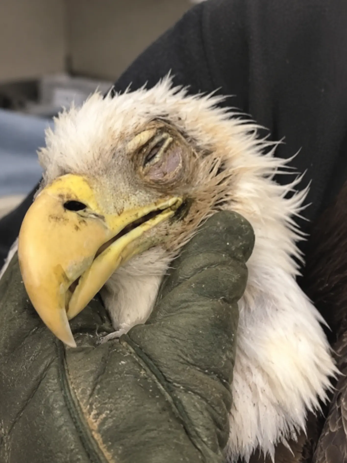
That was the first clue that this adult Bald Eagle might not be dealing with the kind of roadside trauma rescuers initially expected.
The eagle had been found sitting low beside a rural road near the Upper Mississippi River in Wisconsin. It was still conscious, but it did not attempt to fly when people approached from a distance. Its posture was weak, its movements were slow, and one eye was markedly swollen and nearly closed.
Because the bird was found so close to traffic, a vehicle strike seemed like the most likely explanation.
Bald Eagles regularly feed on road-killed animals, particularly during colder months when carrion can provide an easy source of calories. Unfortunately, feeding beside roads places them directly in the path of passing vehicles.
But when the wildlife officer reached the scene and examined the eagle more closely, several expected signs were missing.
There was no obviously broken wing.
No major open wound.
No severe bleeding.
No clear deformity suggesting a catastrophic fracture.
Yet the eagle looked far too sick to be left where it was.
“We took it to a veterinarian, and after the initial examination they transferred the eagle to our team for specialized care,” the officer said.
What followed became less a straightforward trauma case and more a diagnostic search for a hidden cause.
The Eagle Was Found Where a Vehicle Strike Seemed Obvious
The first report came from someone traveling along a rural road who noticed the Bald Eagle remaining unusually close to the shoulder.
At first, the bird’s location did not seem extraordinary.
Bald Eagles commonly move between rivers, farmland, wooded corridors, and roadsides. They may descend to feed on fish, carrion, or other readily available food.
But healthy adult eagles rarely remain on the ground when humans or vehicles approach closely.
This bird did.
It stayed low.
Its wings remained folded.
Its head periodically dropped.
One eye appeared abnormal even from a distance.
When the eagle failed to fly after being given sufficient space, the observer contacted wildlife authorities.
That decision may have been critical.
A severely debilitated eagle can deteriorate quickly, particularly when exposed to cold, traffic, predators, or additional stress.
The Wildlife Officer Expected Trauma
Roadside Bald Eagle calls often raise immediate concern for vehicle collisions.
The reasoning is straightforward.
Eagles are scavengers.
They frequently feed on deer, raccoons, and other animals killed by vehicles.
When a car approaches, a large eagle may not be able to gain altitude quickly enough to clear traffic, especially after feeding heavily.
For that reason, the responding officer approached this bird expecting to find evidence of impact.
Instead, the examination at the scene created more questions.
The eagle was weak.
Its feathers were somewhat disheveled.
Its reactions were delayed.
One eye showed obvious swelling and inflammation.
But the wings were not hanging at dramatically different angles.
There was no visible open fracture.
The legs were present in normal position.
There was no catastrophic external injury that immediately explained the bird’s inability to fly.
“There was no obvious broken wing or major external wound,” the officer said. “But it looked terrible. It was weak, slow to respond, and that eye was badly swollen.”
That changed the rescue plan.
A Bird That Looks Calm May Actually Be Critically Ill
Wildlife rehabilitators often emphasize that a wild raptor allowing close human approach should never automatically be interpreted as calm or cooperative.
A healthy Bald Eagle is typically wary.
It will move away.
It may vocalize.
It may raise its feet defensively.
It may attempt to fly.
This eagle barely resisted.
That lack of resistance was a medical warning.
When a wild predator no longer has the strength to defend itself normally, the underlying illness may already be advanced.
The officer used heavy protective gloves and controlled the bird’s feet first.
Even a weakened eagle can suddenly strike with its talons.
The body was then supported carefully and placed into a secure transport carrier.
No one attempted to force the bird to fly.
There was no value in “testing” an animal that was already demonstrating severe weakness.
The First Veterinarian Found No Major Orthopedic Injury
The eagle was transported to a nearby veterinary clinic for an initial examination.
The veterinary team assessed the wings, shoulders, legs, feet, pelvis, spine, head, and chest.
The bird’s overall condition remained concerning.
It appeared dehydrated.
Grip strength was reduced.
The eagle was quieter than expected.
The affected eye was significantly inflamed.
But the orthopedic examination still did not reveal the kind of major fracture expected after a high-impact vehicle collision.
That was important.
A fracture would have provided a clear mechanical explanation for the inability to fly.
Without one, the team had to consider something broader.
The local veterinarian determined that the eagle required more specialized avian diagnostics and prolonged supportive care than could be provided there.
The bird was transferred to a licensed raptor rehabilitation center.
“No Broken Bone” Did Not Mean “Minor Case”
This is one of the most important parts of the case.
When people hear that no major fracture was found, they may assume the situation was less serious.
In fact, the opposite can be true.
A bird with a visible broken wing has an obvious problem.
A bird with intact wings but profound weakness may be suffering from something that affects the entire body.
Possible causes can include toxin exposure, severe infection, neurological disease, anemia, metabolic abnormalities, dehydration, or other systemic illnesses.
“The absence of a major fracture did not make this a mild case,” the attending veterinarian said. “Something was clearly affecting the eagle far beyond one limb.”
The rehabilitation team therefore began a broader diagnostic workup.
The Eye Injury Became an Important Part of the Examination
Photographs taken during the specialized evaluation show marked swelling and inflammation around one eye.
The eyelids are partially closed.
The surrounding tissue appears irritated.
The eagle remains conscious but subdued while being restrained safely with a protective glove.
An eye that looks this abnormal can have several possible explanations.
There may be direct trauma.
There may be infection.
Inflammation can arise secondary to systemic disease.
A weakened bird may also injure itself after becoming grounded.
Because the cause was not obvious from appearance alone, the team avoided assuming the eye problem explained everything.
Instead, they treated it as one piece of a larger clinical picture.
A Full Ophthalmic Examination Was Necessary
The veterinary team examined the eye for:
corneal injury,
swelling,
abnormal pupil responses,
hemorrhage,
discharge,
and evidence of deeper inflammation.
They also assessed whether the eagle still responded visually from the affected side.
Vision is especially important in a Bald Eagle.
A bird must be able to track movement at distance, judge depth, navigate around obstacles, and land accurately.
Even if the underlying systemic illness resolved, permanent visual impairment could affect release eligibility.
At this stage, however, the priority was stabilization.
Radiographs Helped Rule Out Hidden Trauma
The eagle underwent radiographic imaging to look for injuries that could have been missed during the external examination.
The team examined the wings, chest, shoulders, spine, and other skeletal structures.
They also assessed the head for signs of major impact.
No acute catastrophic fracture was identified.
There was no skeletal injury severe enough to explain the degree of weakness.
That made the original roadside assumption — a simple vehicle strike — less convincing.
It did not make trauma impossible.
A bird can suffer concussion, bruising, or soft-tissue injury without breaking a bone.
But the medical team now had stronger reason to investigate systemic causes.
Bloodwork Became the Next Major Step
Blood was collected to evaluate the eagle’s overall physiological condition.
The veterinary team was interested in several broad questions.
Was the bird anemic?
Was dehydration severe?
Were there signs of inflammation?
Was organ function abnormal?
Was there evidence of a broader infectious process?
The results showed abnormalities consistent with significant systemic illness and dehydration.
Those findings fit what the team was seeing clinically.
The bird looked sick because it was sick.
The question remained why.
Lead Exposure Had to Be Considered
Lead testing is commonly considered in unexplained weakness in Bald Eagles.
These birds are opportunistic scavengers and may ingest fragments of lead ammunition while feeding on animal carcasses or discarded remains.
Lead toxicosis can cause:
weakness,
poor coordination,
reduced grip strength,
gastrointestinal dysfunction,
vision abnormalities,
and inability to fly.
A severely affected eagle may be found on the ground with no broken bones at all.
Because this bird’s weakness could not be explained by trauma alone, lead testing formed part of the broader diagnostic approach.
The team did not assume a positive result in advance.
Testing was used to determine whether lead exposure might be contributing to the clinical signs.
Infectious Disease Also Remained on the List
Another important category was infectious disease.
Certain infections can cause neurological signs, weakness, ocular inflammation, or generalized debilitation.
Samples were collected as appropriate for further evaluation.
Depending on location and clinical signs, wildlife veterinarians may consider diseases such as highly pathogenic avian influenza, West Nile virus, or other infectious conditions.
No single possibility was accepted as the diagnosis simply because it was plausible.
The team continued working from evidence.
Supportive Care Could Not Wait for Every Answer
The eagle was already too compromised to wait for all laboratory results before beginning treatment.
Supportive care started immediately.
Fluids were administered to address dehydration and support circulation.
The bird was housed in a warm, quiet enclosure.
The floor was padded because the eagle was not yet strong enough for normal perching.
Nutritional support was provided cautiously.
The eye was treated according to the findings of the ophthalmic examination.
Pain relief was used when clinically indicated.
The goal during the first phase was not to solve every diagnostic question in a single day.
It was to keep the bird stable long enough for the answers to matter.
The First Night Remained Uncertain
During the first 24 hours, the eagle spent much of its time resting.
Its head remained low.
The affected eye was still partly closed.
It did not consistently maintain an upright posture.
The rehabilitation team minimized handling.
Every examination consumes energy.
For a critically weakened wild bird, unnecessary restraint can create additional physiological stress.
Staff therefore observed as much as possible without disturbing the eagle.
They monitored:
breathing,
posture,
alertness,
grip strength,
food intake,
and neurological responses.
The prognosis remained guarded.