The first sign that something was wrong was not that the Bald Eagle could not fly.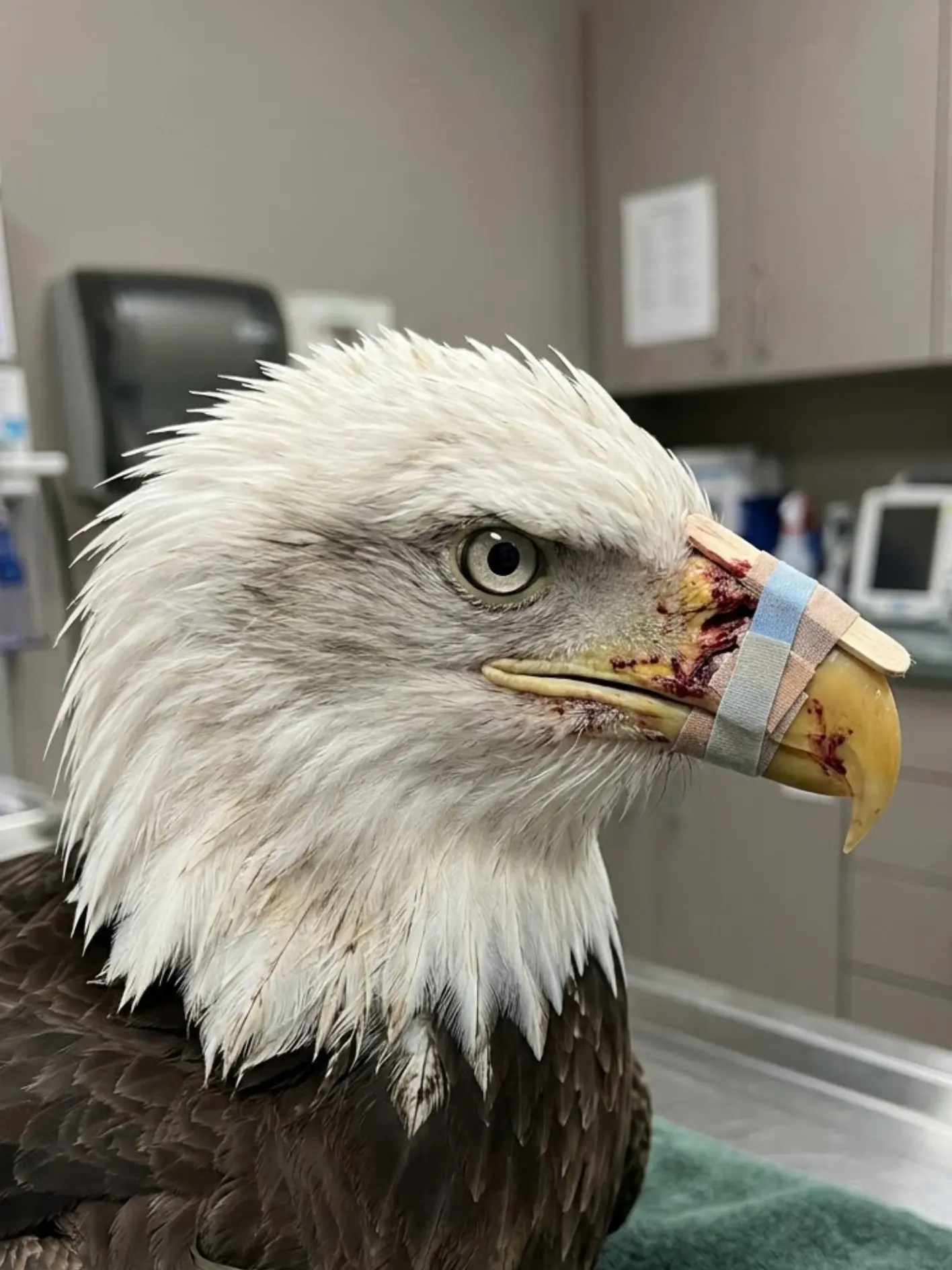
It was that it could not eat.
The adult eagle had been seen near an agricultural area outside Yakima, Washington, repeatedly approaching food on the ground but failing to tear or hold it normally. Each time the bird lowered its head, it appeared to struggle with the same basic movement. It could pick at food, but pieces slipped away. It opened and closed its beak awkwardly. From a distance, observers could see dark staining around the upper bill.
When the eagle finally lifted its head, the reason became clearer.
There was blood around the base of the upper beak.
Part of the structure appeared unstable.
The eagle was still alert and capable of moving, but a bird that cannot use its beak normally cannot survive for long without help.
Wildlife authorities were contacted, and the Bald Eagle was captured and transported to a licensed wildlife rehabilitation hospital for emergency examination.
What veterinarians found beneath the dried blood was far more serious than a superficial cut.
The upper beak had sustained penetrating trauma near its base. The keratin covering was damaged, underlying structures were exposed, and the remaining portion of the bill moved abnormally when the eagle attempted to open and close its mouth.
Diagnostic imaging raised an even more troubling possibility.
The pattern of damage, together with radiopaque material in the injured region, was considered strongly consistent with gunshot trauma.
The bird had not simply chipped its beak.
Something had struck it with enough force to fracture one of the structures it depends on every day to stay alive.
The challenge now was not merely to close the wound.
Veterinarians had to find a way to make the beak functional again.
The Eagle Was Still Flying — But It Was Losing the Ability to Feed
That distinction made the case unusual.
Many injured Bald Eagles are reported because they are grounded.
A broken wing, neurological impairment, poisoning, collision trauma, or extreme weakness may leave a bird unable to take off.
This eagle could still move through the landscape.
Its wings appeared functional.
Its feet remained strong.
Its eyes were alert.
But its beak was failing.
For a Bald Eagle, that can be just as dangerous as losing flight.
The species relies on the beak to tear fish, carrion, and other food into manageable pieces. The feet hold prey or carcass tissue in place while the hooked upper bill applies force. If that upper structure becomes unstable, feeding becomes inefficient or impossible.
A bird may remain visually strong for several days while gradually losing weight.
By the time weakness becomes obvious, the nutritional deficit may already be significant.
That is why the people who reported the bird did not wait for it to collapse.
They noticed abnormal behavior and called for help.
The Initial Examination Confirmed Major Beak Trauma
At intake, the veterinary team first assessed the eagle’s overall condition.
The bird remained responsive.
Its feet gripped strongly.
The wings folded normally.
There was no obvious major orthopedic injury elsewhere.
Attention quickly shifted to the head.
The area around the upper beak showed blood, swelling, and disrupted tissue. The injury was concentrated near the base of the bill, a particularly important region because it contains the living tissues that support the external keratin sheath.
The team examined the mouth carefully.
They checked the alignment between the upper and lower beaks.
They looked at the palate and oral tissues.
They assessed whether the nostrils remained open.
They evaluated the eyes for associated trauma.
Most importantly, they watched how the injured beak behaved when the eagle attempted to close it.
The answer was concerning.
The remaining structure was not rigid.
Part of the upper bill shifted under pressure.
“The major problem was instability,” the attending veterinarian said. “The beak was still there, but it was not behaving like one solid structure anymore.”
That meant every attempt to bite or tear food could move the fracture.
And every movement could delay healing.
Imaging Revealed the Extent of the Damage
Radiographs were performed to determine whether the trauma involved only keratin or extended into the supporting bone.
The images showed deeper structural injury.
Small radiopaque fragments were also present within the damaged region. Their appearance and distribution, combined with the penetrating nature of the injury, were considered strongly consistent with projectile trauma.
The medical team documented the findings carefully.
Because no one witnessed the moment the eagle was injured, staff did not publicly identify who had fired a shot or under what circumstances.
That question belonged to wildlife law enforcement.
The medical record could establish what veterinarians found:
penetrating beak trauma,
fracture and instability,
soft-tissue injury,
and imaging findings compatible with gunfire.
It could not identify the person responsible.
The Beak Is Not Just a Hard Shell
One of the challenges in explaining beak injuries is that people often think of the beak as if it were a horn-like object attached to the face.
It is more complex than that.
The outer surface is largely keratin, but beneath it are living tissues and supporting skeletal structures. The beak grows continuously and is shaped by normal use and wear.
Damage near the tip may sometimes be manageable if the underlying growth structures remain healthy.
Damage near the base can be far more serious.
If tissue responsible for producing new keratin is destroyed, normal regrowth may be compromised.
If underlying bone is fractured, the external beak may lose support.
If alignment changes, the upper and lower bills may no longer meet correctly.
That can create a cascade of problems.
A slightly misaligned beak may wear unevenly.
Uneven wear can worsen deformity.
Deformity can interfere with feeding and preening.
For a wild bird, those small changes can become major survival problems.
The Veterinary Goal Was Function, Not Appearance
The eagle’s beak did not need to look perfect.
It needed to work.
That became the guiding principle of the case.
Veterinarians were not trying to restore an aesthetically flawless bill.
They were trying to preserve enough alignment and strength for the eagle to:
grasp food,
tear meat,
preen feathers,
maintain normal body condition,
and eventually survive without human assistance.
“The goal was functional alignment,” the veterinarian said. “If the bird ended up with a scar or a slightly irregular surface, that was far less important than whether it could feed itself normally.”
The first step was cleaning and stabilizing the injury.
The Damaged Area Was Carefully Treated
The wound was flushed and examined for devitalized tissue and contamination.
Any tissue that clearly could not survive had to be managed conservatively so that healthy structures were preserved wherever possible.
Pain control was essential.
Beak injuries are highly sensitive, and every movement of the bill can create discomfort.
Antimicrobial treatment was used when clinically appropriate because penetrating wounds carry a significant risk of bacterial contamination.
The team also monitored the nostrils and oral cavity to make sure swelling did not compromise breathing or normal mouth function.
But wound care alone would not solve the mechanical problem.
The beak needed support.
Veterinarians Built a Temporary External Brace
The temporary stabilization visible in treatment photographs may look surprisingly simple.
A lightweight supportive piece was positioned along the damaged upper bill and secured externally to help keep the injured structures aligned.
The brace was not intended to replace the eagle’s beak permanently.
Its purpose was to act as a bridge.
It reduced movement across the injured area.
It helped distribute forces during opening and closing.
And it gave the damaged tissue a better opportunity to heal without being repeatedly pulled apart.
The external support had to be light enough that it did not interfere unnecessarily with normal head movement.
It also had to remain secure while avoiding pressure on healthy tissue.
Because the eagle would eventually need to use the beak during feeding, the device could not simply lock the mouth shut.
This was a functional compromise.
Enough stability for healing.
Enough movement for basic use.
The First Several Days Were Focused on Nutrition
Before the eagle could prove whether the temporary repair worked, staff had to keep the bird nutritionally stable.
Food was presented in small, manageable portions.
Large pieces that required heavy tearing were avoided initially.
The team watched how the eagle picked up food.
Could it close the beak?
Could it hold a piece without the injured section shifting dramatically?
Could it swallow normally?
At first, feeding was inefficient.
The eagle attempted to pick up small pieces and dropped some of them.
It adjusted its head position repeatedly.
The team intervened when necessary to make sure the bird maintained adequate intake.
But they also wanted the eagle to begin using the beak independently as soon as it was safe.
A rehabilitation patient cannot be judged only by what humans can help it do.
The bird must ultimately do these things itself.
The First Successful Bite Became a Major Milestone
Several days into treatment, the eagle picked up a small piece of food and held it.
Then it tore.
The movement was careful and slower than normal, but the beak remained stable enough for the bird to complete the action.
It swallowed the food.
Then it repeated the process.
For the veterinary team, that was more important than any improvement in appearance.
“The first time it tore food on its own, that mattered more to us than how the beak looked,” the veterinarian said. “That was evidence that we might actually be preserving useful function.”
From that point forward, feeding became part of rehabilitation rather than merely supportive care.
The eagle was gradually offered food requiring greater manipulation.
The team monitored whether the brace stayed stable and whether the wound reopened.
No major fresh bleeding developed.
The bird’s confidence using the beak slowly improved.
Body Weight Became One of the Most Objective Measures
An eagle can appear alert while slowly losing weight.
That is why the rehabilitation team did not rely only on behavior.
Body weight was monitored.
Muscle condition was assessed.
Food intake was tracked.
If the eagle could not maintain condition independently, that would suggest the beak was not functioning well enough for eventual release.
Fortunately, weight became more stable as feeding improved.
The bird no longer needed the same level of nutritional assistance.
That was another encouraging sign.
The Team Also Watched for New Keratin Growth
The outer beak is constantly renewed.
In a healthy bird, keratin produced near the base gradually moves outward as the beak grows.
After trauma, that process can become unpredictable.
The veterinary team watched the injured region for signs that viable tissue was continuing to produce new keratin.
They looked for:
healthy surface growth,
cracking,
abnormal discoloration,
separation of the keratin layer,
and evidence that part of the beak was losing blood supply.
The appearance changed gradually.
Some damaged material was expected to slough or wear away.
What mattered was whether stronger new tissue could replace it.
Keratin Growth Was Encouraging — But Not a Guarantee
Even when new keratin appears, normal beak formation is not assured.
If the growth bed has been disrupted unevenly, the beak may regenerate with irregular thickness or direction.
That can require continued monitoring.
In some cases, birds with significant beak injuries may need corrective trimming or additional stabilization.
For a permanently captive bird, repeated management may be possible.
For a wild eagle, the standard is much higher.
A release candidate should not require frequent human maintenance simply to keep the beak usable.
That was another reason the team remained cautious.
The question was not whether veterinarians could keep the beak functional inside a hospital.
It was whether the eagle could eventually maintain it naturally.
Preening Became the Next Important Test
Eating is the most obvious use of the beak.
Preening is almost as important.
Birds use the beak to clean and align feathers.
For a Bald Eagle, healthy feathers are essential for flight, insulation, and protection from weather.
An eagle unable to preen normally may gradually develop poor feather condition even if it can eat.
As the injury stabilized, staff began watching the bird during quiet periods.
At first, grooming around the body was limited.
Then the eagle began reaching toward the chest feathers.
Later, it worked through wing feathers with the beak.
Those behaviors were encouraging.
The bird was not simply tolerating the damaged structure.
It was beginning to use it in the normal daily behaviors required for independence.
The Wings Were Never the Main Problem
That made this case very different from many raptor rehabilitation stories.
The eagle did not need weeks of flight-muscle rehabilitation because of a fractured wing.
Its wings were intact.
Once medically stable, flight itself was likely to be one of the easier tests.
The harder question was whether the eagle could feed and maintain itself after release.
“For this bird, flying may be the easy part,” one rehabilitator said. “The real release test is whether that beak can do every job it has to do once no one is preparing food for it.”
That standard shaped the remainder of rehabilitation.
The Bird Became Increasingly Defensive
As strength and feeding improved, the eagle’s behavior changed too.
It became less tolerant of handling.
It watched staff more closely.
It reacted more quickly when someone entered the enclosure.
Its feet became more defensive.
This was welcomed.