An adult Bald Eagle undergoing long-term treatment for a severely damaged beak returned to surgery today for another reconstructive procedure, giving veterinarians their clearest look yet at how the injury is healing beneath the protective acrylic covering.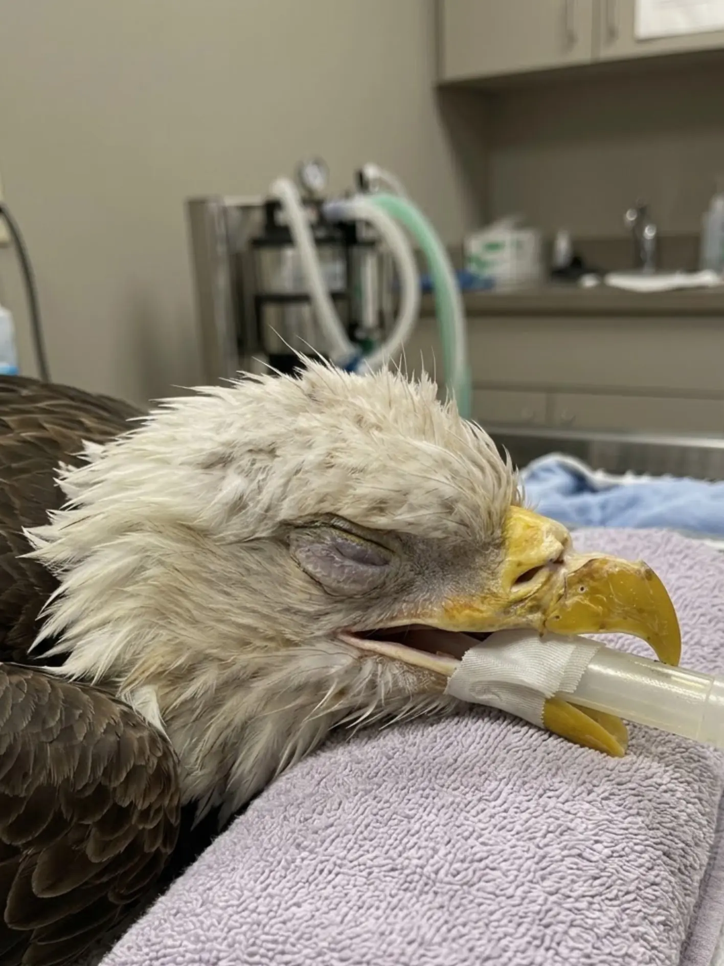
The examination brought both encouraging news and an important reason for continued concern.
The right side of the defect has now filled in completely, allowing the medical team to reduce the amount of acrylic material needed to protect the injured portion of the beak.
The left side, however, remains significantly damaged.
A relatively large gap in the underlying bone is still present, although veterinarians say the area continues to show gradual healing.
And one thing the team had hoped to see by this stage still has not appeared.
New keratin.
Keratin forms the hard outer covering of a Bald Eagle’s beak. It is the visible material that gives the beak its strength, shape and durable cutting surface.
The bone underneath the damaged area appears to be making progress.
The natural keratin sheath has not yet shown clear evidence that it is regenerating.
That distinction has become the central question in the eagle’s recovery.
“We are encouraged by how much the right side has filled in and by the continued healing underneath the remaining defect,” the attending wildlife veterinarian said. “What we are still waiting to see is new keratin. Until that begins to appear, we cannot predict with confidence what the final structure and function of this beak will be.”
For now, the team remains cautiously hopeful.
The tissues still appear capable of healing.
There has been enough improvement to justify continued reconstruction rather than assuming the damage is permanent.
But the longer the injured area remains without visible keratin growth, the greater the concern that the specialized tissues responsible for producing the natural beak covering may have been permanently compromised.
Today’s procedure was therefore about more than replacing an acrylic patch.
It was another opportunity to ask the question that may ultimately determine this eagle’s future:
Can its natural beak begin rebuilding itself?
The Acrylic Covering Is Temporary — Not a Permanent Artificial Beak
When people first see photographs of the eagle’s treatment, the acrylic material can easily look like an artificial replacement for the natural beak.
That is not its purpose.
The acrylic functions primarily as a protective barrier.
Beneath the hard outer keratin sheath, a bird’s beak contains living tissue and bone. When a serious injury removes a large portion of the keratin, the deeper structures can become vulnerable to contamination, trauma, dehydration and additional mechanical damage.
The temporary acrylic covering shields those structures while healing takes place.
It also helps maintain stability around the injured area.
“The acrylic is not intended to replace the natural beak forever,” the veterinarian explained. “Our goal is to protect the tissue underneath long enough to determine how much the eagle can regenerate naturally.”
That makes each replacement procedure an important checkpoint.
The old material is removed.
The underlying defect is inspected.
The team compares what it sees with previous examinations.
Then a new layer is shaped according to how much protection is still necessary.
The reconstruction changes as the eagle changes.
Today’s Procedure Revealed That Part of the Defect Has Closed
The strongest positive finding during the latest examination involved the right side of the original injury.
That portion has now filled in sufficiently that the veterinary team no longer needs to cover as much of the beak with acrylic.
This may sound like a small change.
Clinically, it is significant.
It means at least part of the damaged area has progressed enough that artificial protection can be reduced.
The team was able to apply a smaller acrylic covering during the latest procedure, concentrating protection over the region that remains structurally vulnerable.
“The fact that we can use less material is encouraging,” the veterinarian said. “It means there is less exposed or unsupported area than there was before.”
That reduction also serves another important purpose.
It gives clinicians more natural tissue to observe between procedures.
A Smaller Acrylic Patch Will Make Keratin Easier to Detect
Until now, a larger section of the injured beak has needed artificial coverage.
That protection was necessary, but it also limited direct observation of the healing surface.
With the right side now filled, the new acrylic patch can be smaller.
That means more of the natural margin can remain visible.
If keratin begins emerging along the healing edge, staff may be able to identify it sooner.
“Reducing the coverage gives us a better opportunity to watch what the tissue is doing,” the veterinarian said. “We still have to protect the remaining defect, but we also want to see whether the eagle begins producing its own natural sheath.”
Even a very small amount of new keratin would be meaningful.
The team is not waiting for the beak to suddenly appear normal.
They are looking for a thin margin.
A slight ridge.
A small area of hardened natural material developing along the healing edge.
At this stage, subtle change may be the most important change.
The Left Side Remains the Main Structural Concern
While the right side has shown substantial progress, the left side remains much more complicated.
A sizeable gap in the underlying structure is still present.
The area is not static — the veterinary team says it is gradually filling — but it has not yet healed enough to be left unprotected.
That is why some acrylic remains necessary.
The material provides a protective shell over the vulnerable portion while deeper tissues continue repairing.
The medical team will continue watching the left side for several things:
whether the gap continues to shrink,
whether tissue remains healthy,
whether infection develops,
whether the structure remains stable,
and whether natural keratin begins appearing along the margins.
The size of the remaining defect means this is still a long-term case.
Bone Healing and Keratin Regrowth Are Two Different Problems
One of the most important aspects of this case is that improvement in the underlying bone does not automatically mean the visible beak will regenerate normally.
The beak is a composite structure.
Internally, there is bone and living vascular tissue.
Externally, there is a hard keratin sheath.
Those structures depend on one another, but they do not heal in exactly the same way.
The underlying defect may gradually fill.
Soft tissue may stabilize.
Blood supply may remain adequate.
Yet the natural keratin-producing tissue may still fail to recover completely.
That is why the team is encouraged by the structural healing while remaining cautious about the final outcome.
The eagle is improving.
The beak is not yet proven capable of restoring itself.
Why Keratin Matters So Much
A Bald Eagle’s beak is not merely decorative.
It is a working tool.
The outer keratin layer provides a strong, relatively lightweight surface capable of withstanding repeated mechanical stress.
An eagle uses its beak to:
tear meat,
grasp and reposition food,
remove tissue from carcasses,
preen feathers,
manipulate objects,
and perform normal maintenance behaviors.
The characteristic hooked tip helps the bird tear food efficiently.
If a major section of the keratin sheath fails to regenerate, the underlying structures may remain vulnerable.
The beak could become mechanically weaker.
Its shape might become abnormal.
Wear could become uneven.
The eagle might require repeated artificial reinforcement.
Those possibilities matter greatly when veterinarians begin thinking about long-term welfare and eventual release.
Keratin Is a Living Process Even Though the Material Itself Is Not Living
The hard keratin that forms the outer beak does not contain living cells in the same way deeper tissues do.
But it is continually produced by specialized living tissues.
That is why a healthy bird’s beak can grow and remodel over time.
Normal use causes wear.
Normal growth replaces what is lost.
When trauma destroys part of the structures responsible for producing keratin, that balance can be disrupted.
The question in this case is whether enough of those specialized tissues survived the original injury to begin generating a new natural surface.
The medical team does not yet know.
No Visible Keratin Yet Is Concerning — But Not Definitive
Veterinarians had hoped to see evidence of new keratin by this stage of rehabilitation.
So far, they have not.
That is a concern.
It is not yet a final answer.
Healing rates vary.
The injury was significant.
Tissue that has experienced major trauma may require more time before normal regenerative processes become visible.
The fact that the surrounding area continues to improve gives the team reason to continue waiting.
If the tissue were deteriorating, becoming infected or losing viability, the outlook would be substantially more pessimistic.
Instead, the defect is changing.
The right side has filled.
The left side is gradually improving.
Those developments suggest biological healing is still occurring.
“What we do not want to do is declare the keratin permanently lost simply because it has not appeared as quickly as we hoped,” the veterinarian said. “The underlying area is still making progress, so we believe it is reasonable to continue giving it time.”
Time, However, Is Becoming an Important Part of the Prognosis
Although the team remains hopeful, there is a limit to how long the absence of keratin can remain reassuring.
As more time passes without evidence of regrowth, the possibility of permanent damage becomes increasingly important.
The concern is that the original injury may have destroyed enough of the keratin-producing tissue that complete regeneration will never occur.
If that proves true, the beak may require long-term management.
That could include repeated protective reconstruction or other methods of maintaining a usable surface.
Whether such management would provide an acceptable quality of life would depend on how the eagle functions.
The decision cannot be made yet.
The Eagle Has Already Undergone Multiple Procedures
This latest operation is part of an ongoing reconstructive process rather than a single definitive surgery.
At each procedure, the medical team removes the previous acrylic material and inspects the tissue beneath it.
The process allows veterinarians to evaluate healing directly rather than relying only on external appearance.
Measurements and photographs can be compared with previous examinations.
Areas of improvement become visible.
Areas of concern can be identified before they worsen.
The new acrylic is then shaped according to the current anatomy.
As the natural tissues fill in, the artificial covering can ideally become smaller.
That is exactly what happened today.
Why the Acrylic Cannot Simply Stay in Place Indefinitely
A temporary reconstruction is not something that can simply be applied once and forgotten.
The beak continues changing.
The eagle uses it.
The material experiences wear.
The underlying tissue heals beneath it.
The acrylic can loosen, crack or become poorly fitted as the anatomy changes.
That means periodic replacement is necessary.
More importantly, the veterinary team needs to see what is happening underneath.
Leaving the same covering in place indefinitely would make it difficult to evaluate whether natural tissue is improving or deteriorating.
Each replacement provides another opportunity to inspect the biological repair process.
Anesthesia Allows Precision Without Excessive Stress
The eagle must be safely anesthetized for these detailed procedures.
A Bald Eagle cannot be expected to remain completely motionless while clinicians work millimeters from sensitive structures around the beak and face.
Anesthesia prevents sudden head movement.
It allows the veterinary team to work accurately.
It also reduces the stress associated with prolonged physical restraint.
During the procedure, the bird’s breathing and other physiological parameters are carefully monitored.
Airway support is used as appropriate.
Images taken during this stage can look alarming because the eagle appears completely unresponsive.
That is intentional.
The bird is under controlled veterinary anesthesia while the reconstruction is performed.
The Team Also Watches Closely for Infection
Whenever trauma exposes deeper structures around a beak, infection is an important concern.
Keratin normally acts as a protective barrier.
When that barrier is missing, bacteria can reach tissues that are not designed to remain exposed.
At each recheck, veterinarians evaluate for:
swelling,
abnormal discharge,
odor,