An adult bald eagle being treated at a wildlife rehabilitation center in eastern North Carolina has developed a rapidly worsening anemia, with its packed cell volume — or PCV — eventually falling below 6 percent, leaving veterinarians concerned that the bird’s ability to carry oxygen to its tissues has reached a critically dangerous level.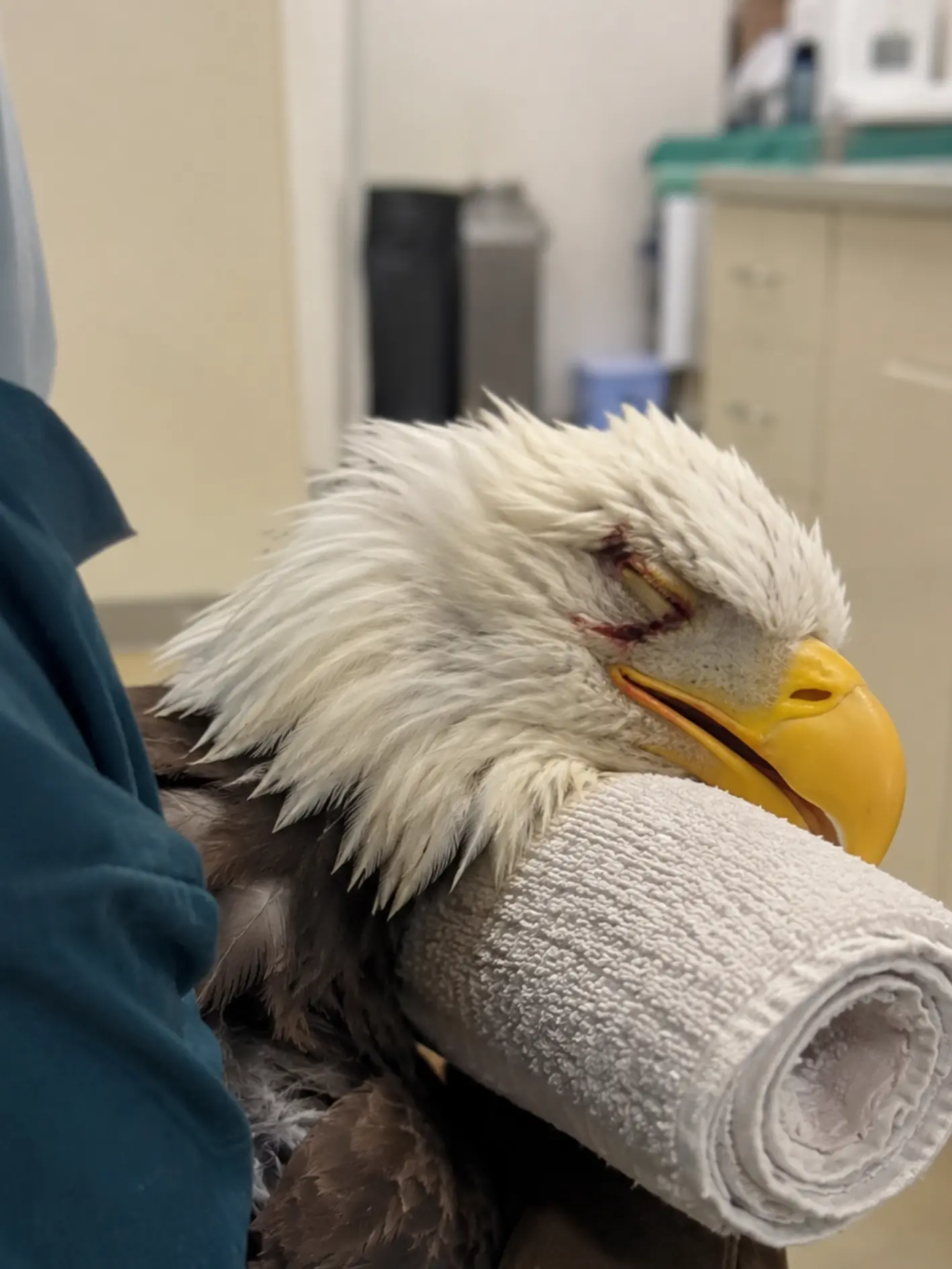
The eagle was originally admitted after being found weak and unable to function normally in Beaufort County, near the Pamlico River. Visible trauma around one eye immediately drew attention during the initial examination, but as the medical team began evaluating the bird more closely, bloodwork revealed a second problem that would quickly become far more urgent.
The eagle was anemic.
At first, veterinarians hoped the abnormal blood value might stabilize with supportive treatment and rest.
It did not.
Repeat testing showed that the percentage of red blood cells in the eagle’s circulation was continuing to decline.
The packed cell volume fell into the single digits.
Then it dropped below 6 percent.
At that point, the medical team was no longer dealing simply with a weak wildlife patient recovering from trauma.
They were dealing with a hematologic emergency.
“The most concerning part was not simply that the eagle was anemic,” a veterinarian involved in the bird’s care said. “It was that the PCV continued to fall despite supportive care. Once it dropped below 6 percent, we were dealing with a patient whose ability to deliver oxygen to its tissues had become critically compromised.”
The wildlife team is now trying to answer the question that will determine nearly every treatment decision that follows:
Why are the eagle’s red blood cells disappearing?
What a PCV Below 6 Percent Means
Packed cell volume is a measurement used to estimate how much of an animal’s circulating blood consists of red blood cells.
Those cells are essential because they carry oxygen from the respiratory system to tissues throughout the body.
When red blood cell volume falls, the blood becomes less capable of delivering adequate oxygen.
The lower the value drops, the harder the body must work to compensate.
Heart rate may increase.
Respiratory effort may change.
Muscles become weak.
The brain and other organs may receive less oxygen than they need.
An animal may remain conscious even while the physiological margin for further decline becomes extremely small.
For a bird of prey, profound anemia is especially concerning because flight is one of the most metabolically demanding forms of locomotion in the animal kingdom.
An eagle with critically reduced oxygen-carrying capacity may be unable to sustain even ordinary activity, let alone the powerful takeoff and prolonged flight required for survival in the wild.
A PCV below 6 percent therefore represented far more than an abnormal laboratory result.
It was evidence that the eagle’s entire body was operating with an extremely limited oxygen reserve.
The Eagle Had Arrived With Visible Trauma
When the adult bald eagle first entered care, the most obvious abnormality was around the head.
One eye was injured, with swelling and visible trauma around the surrounding tissues.
The bird appeared subdued during handling.
The image recorded during treatment shows the eagle being supported by veterinary staff while a towel is positioned under the head and neck. The injured eye appears closed, and there is visible trauma around the periocular region.
At first glance, the case might have appeared to be primarily traumatic.
But the external injury did not explain everything.
The eagle’s weakness seemed disproportionate.
Its energy remained low.
As bloodwork was repeated, the veterinary team began to realize that the more dangerous process was occurring somewhere they could not see.
Inside the bloodstream.
“The eye injury was obvious,” the veterinarian said. “But it did not explain why the red cell volume kept dropping. That is when the case changed from treating visible trauma to trying to determine whether the bird was losing blood, destroying red cells, or failing to replace them.”
The First Blood Test Raised Concern
Initial bloodwork showed that the eagle was already anemic.
At that stage, several explanations remained possible.
The bird may have lost blood before rescue.
Trauma could have produced external or internal bleeding.
Dehydration might have temporarily altered how the initial values appeared.
Systemic illness could have contributed.
The team therefore did what wildlife veterinarians often do when the first laboratory result raises a question but does not yet provide an answer.
They repeated the test.
The hope was that the PCV would remain stable or begin to rise as the eagle received supportive care.
Instead, it continued to fall.
That trend was more alarming than any single number.
One low value can sometimes represent a condition that has already stabilized.
A declining sequence suggests an ongoing process.
Something was still happening.
Was the Eagle Continuing to Lose Blood?
The first major possibility veterinarians considered was active blood loss.
The visible head injury had to be evaluated carefully.
Any external wounds were examined for ongoing bleeding.
The mouth and oral tissues were checked.
Staff looked for blood around the cloaca or in fecal material that might suggest gastrointestinal bleeding.
The body was also examined for signs that hemorrhage could be occurring internally.
In a wild bird, hidden bleeding can be difficult to identify.
Dense feathers conceal bruising.
Fluid may accumulate internally without immediately producing dramatic external signs.
Depending on the bird’s stability and the resources available, radiographs or other imaging can help identify abnormalities that might be consistent with internal trauma.
But the team could not simply assume that bleeding was the answer.
If blood loss alone were responsible, they still needed to determine where it was occurring and whether it was continuing.
Could the Red Cells Be Breaking Down?
The second broad possibility was hemolysis — destruction of red blood cells.
This can occur for multiple reasons.
Certain toxins can interfere with normal red cell survival.
Some infections may damage blood cells directly or indirectly.
Immune-mediated processes can sometimes cause the body to destroy its own circulating cells.
The veterinary team therefore began looking for evidence that the eagle’s blood cells were being destroyed faster than the body could replace them.
Microscopic examination of a blood smear can provide important clues.
Veterinarians can look at the size and shape of red cells.
They can search for abnormal cell destruction.
They can examine whether immature blood cells are appearing in circulation, which may indicate that the body is attempting to regenerate what has been lost.
They can also look for organisms or other abnormalities that might suggest an infectious or parasitic process.
The blood itself becomes part of the investigation.
Was the Bone Marrow Failing to Keep Up?
A third possibility was that the eagle was not producing enough new red blood cells.
Normally, when red cells are lost, the body responds by increasing production.
That regenerative response may become visible in the blood.
If the body fails to respond appropriately, the problem may involve chronic disease, nutritional compromise, bone marrow suppression, or another systemic process interfering with normal blood-cell production.
For the medical team, this distinction matters enormously.
An eagle actively replacing lost red cells may have a better chance of recovery if the underlying cause can be stopped.
An eagle that is not producing replacements faces a more complicated prognosis.
Veterinarians therefore wanted to know not only how few red cells remained, but whether the eagle’s body was trying to rebuild them.
Lead Exposure Was One Possibility
Because the patient was a bald eagle, lead exposure had to be considered.
Bald eagles are opportunistic scavengers.
They may feed on animal remains containing small fragments of ammunition.
Those particles can be ingested unintentionally.
Lead can affect multiple body systems, including the nervous system, gastrointestinal tract, and normal blood-cell processes.
Weakness is common in affected birds.
Neurological abnormalities may occur.
Anemia can also become part of the clinical picture.
For that reason, blood lead testing was considered an important part of the diagnostic workup.
But the team was careful not to treat lead as a confirmed explanation.
The bird’s species and symptoms made it a reasonable differential diagnosis.
They did not make it a diagnosis.
“Lead is something we absolutely have to consider in a bald eagle,” the veterinarian said. “But we cannot look at a falling PCV and simply decide that lead is the answer. We need testing, and we need to continue looking for other causes at the same time.”
Rodenticide Exposure Was Also Considered
Another possibility involved rodenticides.
Some anticoagulant rodenticides interfere with normal blood clotting.
A predator or scavenger can potentially be exposed indirectly after consuming contaminated prey.
If clotting becomes severely impaired, internal or external bleeding may occur.
That could produce progressive anemia.
But rodenticide exposure cannot be diagnosed from anemia alone.
The veterinary team would need supporting evidence from the clinical picture, laboratory testing, coagulation assessment, or toxicology.
Other types of rodenticides work through different mechanisms and may not produce the same signs.
For that reason, “rodenticide poisoning” could not be used as a blanket explanation.
It remained one possibility among several.
Parasites Could Not Be Ignored
Parasites also entered the differential diagnosis.
Wild birds frequently carry some parasites without becoming critically ill.
But heavy parasite burdens can contribute to chronic blood loss, poor condition, or systemic stress.
Blood parasites may directly affect circulating blood cells.
Intestinal parasites can interfere with nutrition or contribute to ongoing loss depending on the species and severity of infection.
The team therefore considered fecal examination, blood smear evaluation, and other parasite screening as part of the broader investigation.
Again, the goal was not to attach every abnormality to a single cause prematurely.
The eagle could theoretically be dealing with more than one problem at the same time.
That is common in wildlife medicine.
The Visible Eye Injury Might Have Been Only One Piece
The injury around the eagle’s eye remained medically relevant.
It required treatment and monitoring.
But veterinarians became increasingly reluctant to assume it was the sole explanation for the anemia.
The amount of visible blood did not appear sufficient to explain a PCV that continued falling to below 6 percent.
That raised the possibility of either hidden hemorrhage or a completely separate systemic process.
The photograph of the eagle during treatment captures that contrast well.
The external injury is immediately visible.
The life-threatening anemia is not.
A viewer can see the damaged eye.
No one can see a critically low red blood cell volume without laboratory testing.
That is why repeated bloodwork changed the direction of the case.
Handling Became More Dangerous as the PCV Fell
As the eagle became more severely anemic, every procedure had to be considered carefully.
Handling itself causes stress.
Stress increases oxygen demand.
A healthy bird can usually compensate.
A bird with a PCV below 6 percent has far less physiological reserve.
The team therefore minimized unnecessary restraint.
The eagle was kept quiet.
Environmental stimulation was reduced.
Body temperature was carefully supported.
When supplemental oxygen was considered clinically appropriate, it could help increase the amount of oxygen available to the bird even though it could not replace the missing red cells.
Oxygen therapy does not correct anemia.
It supports the patient while the underlying problem is being addressed.
Fluid Therapy Had to Be Balanced Carefully
Hydration presented another clinical challenge.
A sick eagle may require fluid support.
Dehydration can worsen circulation and complicate recovery.
But excessive fluid administration can dilute the blood further.
When the red-cell concentration is already critically low, that matters.
The team therefore had to balance two competing needs:
support circulation without causing unnecessary hemodilution.
“Fluid therapy had to become much more deliberate once the PCV reached that level,” the veterinarian said. “We still needed to support the bird, but we also had to be very conscious that an already critically low red-cell concentration could be diluted further.”
This is one reason severe anemia turns seemingly routine supportive care into a more complex medical decision.
Even Blood Collection Had to Be Limited
One of the paradoxes of the case was that clinicians needed repeated blood testing to understand what was happening.
But every blood sample removes some blood from the patient.
In a healthy eagle, a small diagnostic sample is usually well tolerated.
In a bird with a PCV below 6 percent, unnecessary blood loss becomes unacceptable.
Sample size therefore had to be carefully controlled.
Tests had to be prioritized.
The team could not repeatedly draw large volumes simply because additional information might be useful.
Once the PCV fell into the single digits, every diagnostic blood collection had to be weighed against the fact that the patient was already carrying dangerously few red cells.
That meant relying heavily on trends, clinical signs, and carefully selected testing.
The Team Watched More Than the Number
PCV was central to the case, but it was not the only measure of the eagle’s condition.
Staff monitored breathing.
Heart and circulatory stability.
Mental alertness.
Ability to remain upright.
Appetite.
Body temperature.
Response to handling.
Color of visible tissues.
Overall weakness.
An eagle whose PCV remains low but becomes more alert may be compensating.
A bird whose laboratory number changes only slightly while respiratory effort and weakness worsen may be clinically deteriorating.
Veterinarians therefore interpreted the PCV alongside the entire patient.
The number matters because it quantifies the anemia.
The bird matters because it reveals whether the body is tolerating it.
A Blood Transfusion Became a Serious Consideration
Once the PCV fell below 6 percent, blood transfusion became one of the options the veterinary team had to consider if the eagle continued declining or showed worsening evidence that its tissues were not receiving enough oxygen.
Transfusion in avian wildlife is not a routine procedure.
A suitable donor has to be available.
Blood must be collected safely.
The amount administered has to be calculated carefully.
The recipient has to tolerate the procedure.
Logistical challenges can be substantial, especially at a wildlife rehabilitation facility without immediate access to multiple healthy large raptors.
But when anemia becomes profound enough, transfusion can provide something no medication can produce instantly:
additional circulating red blood cells.
That can increase oxygen-carrying capacity while the medical team continues trying to stop the process causing the anemia.
A Transfusion Would Buy Time, Not Solve the Case
Even if the eagle received donor blood, the underlying problem would remain.
If active hemorrhage continued, the transfused cells could be lost.
If red cells were being destroyed, donor cells might also decline.
If bone marrow production was impaired, the bird would still need treatment for the cause of that suppression.
A transfusion therefore cannot be thought of as a cure.
It is a bridge.
It may provide enough physiological support to keep the eagle alive while diagnostics and treatment catch up.
That distinction is critical.
“We can support the bird,” the veterinarian said, “but unless we understand why those red cells are being lost or destroyed, we are treating the consequence rather than the disease.”
Nutrition Became Part of the Hematologic Treatment
The eagle also needed adequate nutritional support.
Red blood-cell production requires energy and nutrients.
Healing from trauma requires protein.
An animal struggling with severe anemia may lose appetite because simply maintaining normal activity becomes difficult.
Staff therefore monitored how much the eagle was eating and whether supplemental nutritional support was necessary.
The objective was not to treat a PCV of less than 6 percent with food alone.
Nutrition cannot reverse profound anemia overnight.
But without sufficient calories and protein, the body has even less ability to produce new cells or repair injured tissues.
The eagle needed every available physiological advantage.
Body Condition Offered Another Clue
Veterinarians evaluated the eagle’s body weight and muscle condition repeatedly.
A markedly thin bird might suggest a longer period of illness before rescue.
A bird in good condition with sudden severe anemia might point clinicians toward a more acute process.
Neither pattern is diagnostic by itself.
But physical condition can help construct a timeline.
Was this an eagle that had been declining for weeks?
Or had something happened recently that caused a rapid loss of red cells?
Those questions could influence which tests were prioritized.
Wildlife medicine rarely provides a complete history.