When the adult Bald Eagle arrived at the wildlife hospital, it no longer had the strength to remain standing. Staff eased the bird onto thick, folded towels inside a quiet treatment enclosure and began the kind of emergency assessment that often follows a serious trauma case. From the outside, the eagle looked profoundly weak, but there was no obvious catastrophic fracture, no active hemorrhage, and no single dramatic wound that fully explained its condition. The first major clue came only after diagnostic testing. Bloodwork revealed that the eagle was suffering from severe lead exposure, a toxic condition that can quietly overwhelm multiple organ systems and leave even a large raptor too weak to fly, hunt, or defend itself.
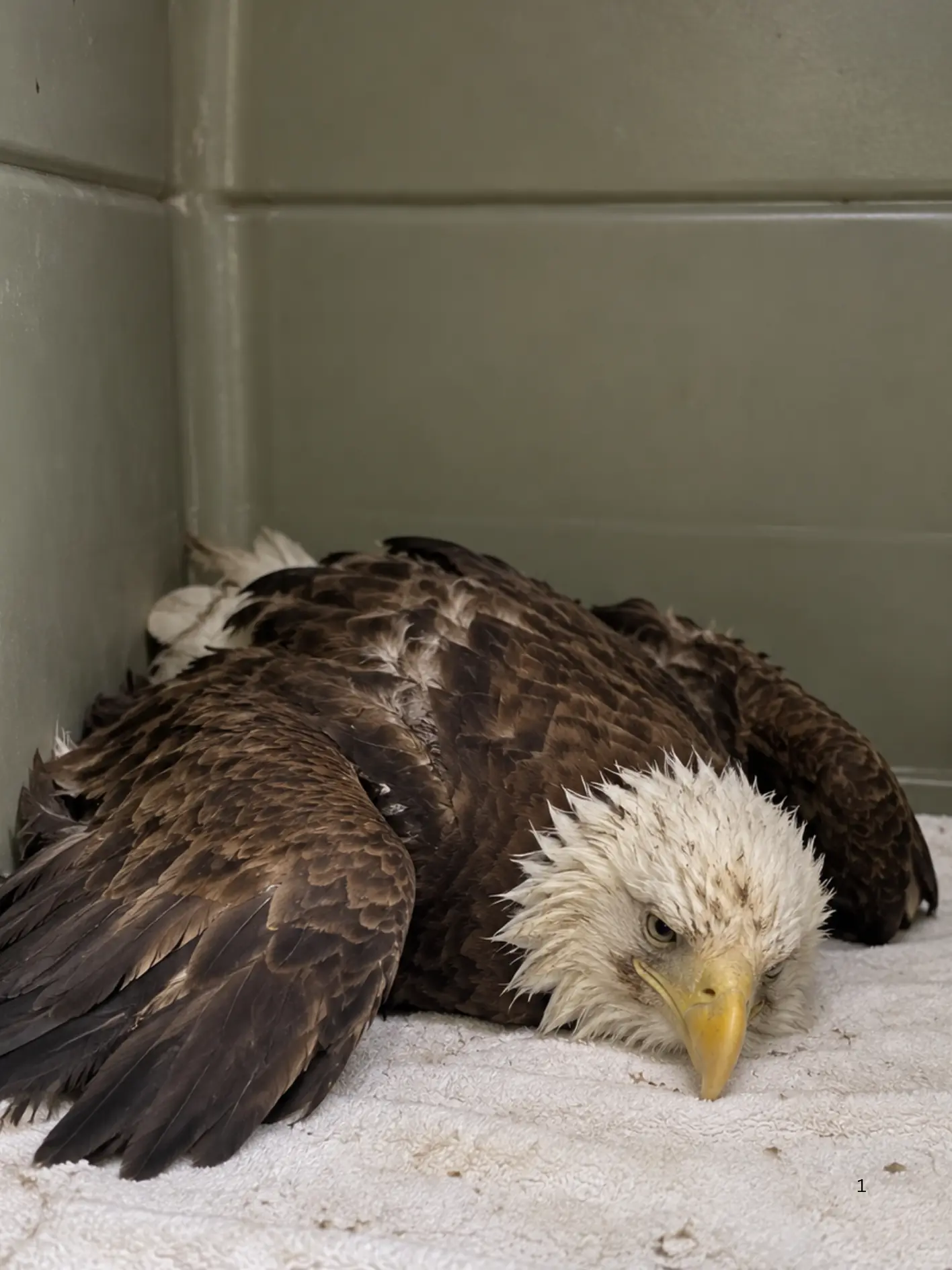
The eagle had been found earlier that day near a small lakeside road in northern Wisconsin. A resident reported seeing the bird standing low to the ground near the edge of the property, with its head held unusually low and its wings drooping. At first glance, the resident thought the eagle might simply be resting. But as time passed, it became clear that something was wrong. The bird did not flush when approached at a distance, which is abnormal for a healthy adult Bald Eagle. Instead, it made a weak effort to move, took a few unsteady steps, and then lowered itself again. It never attempted a sustained takeoff.
That limited response immediately raised concern. Adult Bald Eagles are powerful, alert birds that usually avoid close human approach unless they are severely compromised. In this case, the bird’s tolerance of people was not a sign of calm behavior. It was a sign that it was running out of strength.
A wildlife responder was contacted, and the eagle was professionally captured and transported to a rehabilitation center for emergency care. Even during rescue, the bird’s weakness remained obvious. It still had awareness and could track movement with its eyes, but its grip strength was poor, its posture was unstable, and it offered far less resistance than would be expected from a healthy adult eagle. For experienced responders, that can be one of the most alarming findings. A large raptor that cannot mount a strong defensive response is often in serious systemic trouble.
At intake, veterinary staff began with a full physical examination. The eagle was dehydrated, lethargic, and underpowered. Its posture suggested profound fatigue rather than a purely orthopedic injury. The talons could still flex, but grip strength was reduced. The wings moved symmetrically enough that a severe open wing fracture seemed less likely at first glance, though pain and weakness were clearly present. There was no major external wound that explained the degree of collapse. The bird’s mucous membranes were pale, suggesting poor perfusion or anemia, and its body condition was somewhat reduced. Staff also noted that the eagle’s responses seemed slowed, a finding that can be associated with toxic exposure, metabolic compromise, or neurologic disease.
Because lead intoxication is a well-known cause of weakness in Bald Eagles and other scavenging raptors, the team included it early in the differential diagnosis. Still, trauma could not be ruled out without imaging. Radiographs were performed to assess the skeleton, body cavities, and gastrointestinal tract.
The X-rays did not show a major fracture that would explain the eagle’s inability to fly or stand. That absence of a devastating skeletal injury redirected attention toward medical causes. More importantly, the radiographs revealed multiple tiny metallic-density fragments within the gastrointestinal tract. Those particles were small, but they mattered. Metallic fragments in a scavenging raptor can raise immediate concern for lead exposure, especially when the clinical signs include weakness, poor coordination, gastrointestinal slowdown, and general collapse.
The team then performed in-house screening to evaluate the bird’s blood lead status. The result was deeply concerning. The lead level was above the range that could be precisely measured by the initial analyzer, indicating a heavy toxic burden. A confirmatory sample was sent to a diagnostic laboratory for formal quantification, but the clinical picture had already become clear: the emergency was not a broken bone. It was in the eagle’s bloodstream.
Additional bloodwork supported the seriousness of the case. The eagle showed evidence consistent with systemic compromise, including dehydration and changes suggestive of anemia. Depending on the stage and severity of poisoning, lead can interfere with red blood cell production, damage the gastrointestinal tract, alter neurologic function, and place stress on the kidneys and other organs. In large raptors, the condition can progress to profound weakness, inability to stand, seizures, paralysis, and death if it is not recognized and treated in time.
Lead poisoning in Bald Eagles most commonly results from ingestion of lead fragments. These birds are opportunistic scavengers as well as hunters, and they may feed on carcasses or discarded animal remains that contain tiny pieces of spent ammunition. In some cases, lead fishing tackle can also be a source. Once swallowed, even small fragments can be exposed to the acidic environment of the digestive tract. Over time, that lead can dissolve and enter the bloodstream. The fragments do not have to be large to become dangerous. Pieces that are difficult to see with the naked eye may still be biologically significant.
In this case, because metallic fragments were visible in the digestive tract and the eagle had been found in a region where outdoor recreational activity and hunting occur, ingestion of lead fragments was considered a leading possibility. Still, the rehabilitation team remained careful in its language. The presence of lead toxicity could be confirmed. The exact source of the lead could not.
Treatment began immediately. The eagle was admitted to a quiet, padded enclosure to minimize stress and reduce the risk of further injury. Supportive care was critical from the outset. Fluids were administered to address dehydration and support circulation. Body temperature was monitored closely. Staff limited unnecessary stimulation, as exhausted raptors can deteriorate quickly when handled excessively. Pain management and gastrointestinal support were also incorporated as indicated by the bird’s condition.
The central treatment for the lead toxicity itself was chelation therapy under veterinary supervision. Chelation agents work by binding circulating lead so that the body can excrete it more effectively. This is not a simple or casual intervention. It can be physically demanding on an already compromised patient, and it requires careful monitoring. But in a case of severe lead exposure, it can be lifesaving.
As the attending veterinarian later explained, chelation does not instantly reverse what the toxin has done. It helps reduce the lead burden while the medical team supports the organs and body systems that have already been affected. In other words, the therapy creates a chance for recovery, but it does not guarantee it.
The first 24 to 48 hours were the most concerning. For much of the first night, the eagle remained down on the towels, barely lifting its head. It was conscious and aware, but even maintaining an upright posture seemed difficult. Staff monitored respiratory effort, hydration status, mentation, and neurologic function. Appetite was poor at first, which was not unexpected. Birds with significant lead toxicity often show reduced gastrointestinal motility and a general reluctance or inability to eat normally.
The confirmatory laboratory result later returned and verified severe lead intoxication. That finding aligned with the imaging and the bird’s clinical signs. With the diagnosis confirmed, the treatment plan remained focused on stabilization, chelation, serial monitoring, and gradual nutritional support.
One of the most important parts of managing a case like this is patience. Even when the lead burden begins to decline, the patient may still appear critically weak because the damage caused by the toxin does not disappear overnight. Nerves, muscles, kidneys, gastrointestinal function, and red blood cell production all need time to recover. For the veterinary team, improvement is often measured first in small ways.